 Movie
Movie Controller

[English] 日本語
 Yorodumi
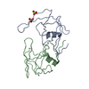
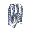
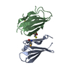
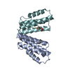
- PDB-2hvp: THREE-DIMENSIONAL STRUCTURE OF ASPARTYL PROTEASE FROM HUMAN IMMUN... -
+ Open data

- Basic information
| Entry | Database: PDB / ID: 2hvp | ||||||
|---|---|---|---|---|---|---|---|
| Title | THREE-DIMENSIONAL STRUCTURE OF ASPARTYL PROTEASE FROM HUMAN IMMUNODEFICIENCY VIRUS HIV-1 | ||||||
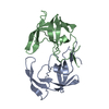
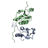
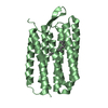
 Components Components | HIV-1 PROTEASE | ||||||
 Keywords Keywords | HYDROLASE(ACID PROTEINASE) | ||||||
| Function / homology |  Function and homology information Function and homology informationHIV-1 retropepsin / symbiont-mediated activation of host apoptosis / retroviral ribonuclease H / exoribonuclease H / exoribonuclease H activity / DNA integration / viral genome integration into host DNA / establishment of integrated proviral latency / RNA-directed DNA polymerase / RNA stem-loop binding ...HIV-1 retropepsin / symbiont-mediated activation of host apoptosis / retroviral ribonuclease H / exoribonuclease H / exoribonuclease H activity / DNA integration / viral genome integration into host DNA / establishment of integrated proviral latency / RNA-directed DNA polymerase / RNA stem-loop binding / viral penetration into host nucleus / host multivesicular body / RNA-directed DNA polymerase activity / RNA-DNA hybrid ribonuclease activity / Transferases; Transferring phosphorus-containing groups; Nucleotidyltransferases / host cell / viral nucleocapsid / DNA recombination / DNA-directed DNA polymerase / aspartic-type endopeptidase activity / Hydrolases; Acting on ester bonds / DNA-directed DNA polymerase activity / symbiont-mediated suppression of host gene expression / viral translational frameshifting / symbiont entry into host cell / lipid binding / host cell nucleus / host cell plasma membrane / virion membrane / structural molecule activity / proteolysis / DNA binding / zinc ion binding Similarity search - Function | ||||||
| Biological species |   Human immunodeficiency virus 1 Human immunodeficiency virus 1 | ||||||
| Method |  X-RAY DIFFRACTION / Resolution: 3 Å X-RAY DIFFRACTION / Resolution: 3 Å | ||||||
 Authors Authors | Navia, M.A. / Fitzgerald, P.M.D. / Mckeever, B.M. / Springer, J.P. | ||||||
 Citation Citation | Journal: Nature / Year: 1989 Title: Three-dimensional structure of aspartyl protease from human immunodeficiency virus HIV-1. Authors: Navia, M.A. / Fitzgerald, P.M. / McKeever, B.M. / Leu, C.T. / Heimbach, J.C. / Herber, W.K. / Sigal, I.S. / Darke, P.L. / Springer, J.P. #1: Journal: J.Biol.Chem. / Year: 1989Title: Crystallization of the Aspartylprotease from the Human Immunodeficiency Virus, HIV-1 Authors: Mckeever, B.M. / Navia, M.A. / Fitzgerald, P.M.D. / Springer, J.P. / Leu, C.-T. / Heimbach, J.C. / Herber, W.K. / Sigal, I.S. / Darke, P.L. | ||||||
| History |
|
- Structure visualization
| Structure viewer | Molecule: MolmilJmol/JSmol |
|---|
- Downloads & links
-Download
| PDBx/mmCIF format | 2hvp.cif.gz | 12.6 KB | Display | PDBx/mmCIF format |
|---|---|---|---|---|
| PDB format | pdb2hvp.ent.gz | 5.3 KB | Display | PDB format |
| PDBx/mmJSON format | 2hvp.json.gz | Tree view | PDBx/mmJSON format | |
| Others |  Other downloads Other downloads |
-Validation report
| Arichive directory | https://data.pdbj.org/pub/pdb/validation_reports/hv/2hvpftp://data.pdbj.org/pub/pdb/validation_reports/hv/2hvp | HTTPS FTP |
|---|
-Related structure data
| Similar structure data |
|---|

-Links
 PDBj
PDBj
- Assembly
| Deposited unit | 
| ||||||||
|---|---|---|---|---|---|---|---|---|---|
| 1 | 
| ||||||||
| Unit cell |
| ||||||||
| Details | THE HIV-1 PROTEASE IS A DIMER. IN THE CRYSTAL THE TWO MONOMERS ARE RELATED BY A CRYSTALLOGRAPHIC TWO-FOLD AXIS. TO GENERATE THE SYMMETRY RELATED MONOMER, THE FOLLOWING TRANSFORMATION MUST BE APPLIED TO THE COORDINATES PRESENTED IN THIS ENTRY 0.0 -1.0 0.0 50.29 -1.0 0.0 0.0 50.29 0.0 0.0 -1.0 53.40 |
-Components
| #1: Protein | Mass: 10830.781 Da / Num. of mol.: 1 Source method: isolated from a genetically manipulated source Source: (gene. exp.) Human immunodeficiency virus 1 / Genus: Lentivirus / References: UniProt: P12497 |
|---|
-Experimental details
-Experiment
| Experiment | Method: X-RAY DIFFRACTION |
|---|
- Sample preparation
| Crystal | Density Matthews: 3.12 Å3/Da / Density % sol: 60.52 % | ||||||||||||||||||||||||||||||||||||||||||
|---|---|---|---|---|---|---|---|---|---|---|---|---|---|---|---|---|---|---|---|---|---|---|---|---|---|---|---|---|---|---|---|---|---|---|---|---|---|---|---|---|---|---|---|
| Crystal grow | *PLUS Temperature: 4 ℃ / pH: 7 / Method: vapor diffusion, hanging drop / Details: referred to J.Biol.Chem. 264,1919-1921 | ||||||||||||||||||||||||||||||||||||||||||
| Components of the solutions | *PLUS
|
-Data collection
| Reflection | *PLUS Highest resolution: 3 Å / Rmerge(I) obs: 0.063 |
|---|
- Processing
| Software | Name: CORELS / Classification: refinement | ||||||||||||
|---|---|---|---|---|---|---|---|---|---|---|---|---|---|
| Refinement | Highest resolution: 3 Å | ||||||||||||
| Refinement step | Cycle: LAST / Highest resolution: 3 Å /
| ||||||||||||
| Refinement | *PLUS Highest resolution: 3 Å / Rfactor obs: 0.37 | ||||||||||||
| Solvent computation | *PLUS | ||||||||||||
| Displacement parameters | *PLUS |
