 Movie
Movie Controller

[English] 日本語
 Yorodumi
- PDB-3uhl: Crystal Structure of Multidrug Resistant HIV-1 Protease Clinical ... -
+ Open data

- Basic information
| Entry | Database: PDB / ID: 3uhl | ||||||
|---|---|---|---|---|---|---|---|
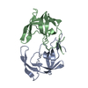
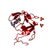
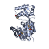
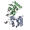
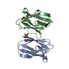
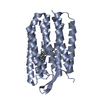
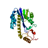
| Title | Crystal Structure of Multidrug Resistant HIV-1 Protease Clinical Isolate PR20 in Complex with p2-NC substrate analog | ||||||
 Components Components | Protease | ||||||
 Keywords Keywords | Hydrolase/Hydrolase inhibitor / Hydrolase-Hydrolase inhibitor complex | ||||||
| Function / homology |  Function and homology information Function and homology informationHIV-1 retropepsin / symbiont-mediated activation of host apoptosis / retroviral ribonuclease H / exoribonuclease H / exoribonuclease H activity / DNA integration / viral genome integration into host DNA / establishment of integrated proviral latency / RNA-directed DNA polymerase / RNA stem-loop binding ...HIV-1 retropepsin / symbiont-mediated activation of host apoptosis / retroviral ribonuclease H / exoribonuclease H / exoribonuclease H activity / DNA integration / viral genome integration into host DNA / establishment of integrated proviral latency / RNA-directed DNA polymerase / RNA stem-loop binding / viral penetration into host nucleus / host multivesicular body / RNA-directed DNA polymerase activity / RNA-DNA hybrid ribonuclease activity / Transferases; Transferring phosphorus-containing groups; Nucleotidyltransferases / host cell / viral nucleocapsid / DNA recombination / DNA-directed DNA polymerase / aspartic-type endopeptidase activity / Hydrolases; Acting on ester bonds / DNA-directed DNA polymerase activity / symbiont-mediated suppression of host gene expression / viral translational frameshifting / symbiont entry into host cell / lipid binding / host cell nucleus / host cell plasma membrane / virion membrane / structural molecule activity / proteolysis / DNA binding / zinc ion binding Similarity search - Function | ||||||
| Biological species |   Human immunodeficiency virus 1 Human immunodeficiency virus 1 | ||||||
| Method |  X-RAY DIFFRACTION / SYNCHROTRON / MOLECULAR REPLACEMENT / Resolution: 2.2 Å X-RAY DIFFRACTION / SYNCHROTRON / MOLECULAR REPLACEMENT / Resolution: 2.2 Å | ||||||
 Authors Authors | Agniswamy, J. / Chen-Hsiang, S. / Weber, I. | ||||||
 Citation Citation | Journal: Biochemistry / Year: 2012 Title: HIV-1 protease with 20 mutations exhibits extreme resistance to clinical inhibitors through coordinated structural rearrangements. Authors: Agniswamy, J. / Shen, C.H. / Aniana, A. / Sayer, J.M. / Louis, J.M. / Weber, I.T. | ||||||
| History |
|
- Structure visualization
| Structure viewer | Molecule: MolmilJmol/JSmol |
|---|
- Downloads & links
-Download
| PDBx/mmCIF format | 3uhl.cif.gz | 173.3 KB | Display | PDBx/mmCIF format |
|---|---|---|---|---|
| PDB format | pdb3uhl.ent.gz | 139.8 KB | Display | PDB format |
| PDBx/mmJSON format | 3uhl.json.gz | Tree view | PDBx/mmJSON format | |
| Others |  Other downloads Other downloads |
-Validation report
| Arichive directory | https://data.pdbj.org/pub/pdb/validation_reports/uh/3uhlftp://data.pdbj.org/pub/pdb/validation_reports/uh/3uhl | HTTPS FTP |
|---|
-Related structure data
| Related structure data | 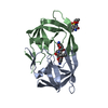 3ucbC 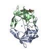 3uf3C 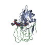 3ufnC 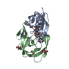 2aodS C: citing same article ( S: Starting model for refinement |
|---|---|
| Similar structure data |


-Links
 PDBj
PDBj


- Assembly
| Deposited unit | 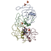
| ||||||||
|---|---|---|---|---|---|---|---|---|---|
| 1 | 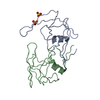
| ||||||||
| 2 | 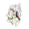
| ||||||||
| Unit cell |
| ||||||||
| Components on special symmetry positions |
|
-Components
| #1: Protein | Mass: 10766.542 Da / Num. of mol.: 4 / Fragment: unp residues 501-599 Mutation: Q7K,L10F,I13V,I15V,D30N,V32I,L33F,E35D,M36I,S37N,I47V,I54L,Q58E,I62V,L63P,A71V,I84V,N88D,L89T,L90M Source method: isolated from a genetically manipulated source Source: (gene. exp.) Human immunodeficiency virus 1 / Gene: pol / Plasmid: pET11a / Production host:  #2: Chemical | ChemComp-SO4 /   Mass: 96.063 Da / Num. of mol.: 4 / Source method: obtained synthetically / Formula: SO4 Mass: 96.063 Da / Num. of mol.: 4 / Source method: obtained synthetically / Formula: SO4#3: Chemical | ChemComp-2NC / |   Type: peptide-like, Peptide-like / Class: Inhibitor / Mass: 770.983 Da / Num. of mol.: 1 / Source method: obtained synthetically / Formula: C35H68N11O8 Type: peptide-like, Peptide-like / Class: Inhibitor / Mass: 770.983 Da / Num. of mol.: 1 / Source method: obtained synthetically / Formula: C35H68N11O8References: N-{(2S)-2-[(N-acetyl-L-threonyl-L-isoleucyl)amino]hexyl}-L-norleucyl-L-glutaminyl-N~5~-[amino(iminio)methyl]-L-ornithinamide #4: Water | ChemComp-HOH / |  Mass: 18.015 Da / Num. of mol.: 106 / Source method: isolated from a natural source / Formula: H2O Mass: 18.015 Da / Num. of mol.: 106 / Source method: isolated from a natural source / Formula: H2O |
|---|
-Experimental details
-Experiment
| Experiment | Method: X-RAY DIFFRACTION / Number of used crystals: 1 |
|---|
- Sample preparation
| Crystal | Density Matthews: 2.52 Å3/Da / Density % sol: 51.26 % |
|---|---|
| Crystal grow | Temperature: 298 K / Method: vapor diffusion, hanging drop / pH: 4.5 Details: 0.9M Ammonium Sulfate, 0.1M Sodium Citrate, pH 4.5, VAPOR DIFFUSION, HANGING DROP, temperature 298K |
-Data collection
| Diffraction | Mean temperature: 100 K | |||||||||||||||||||||||||||||||||||
|---|---|---|---|---|---|---|---|---|---|---|---|---|---|---|---|---|---|---|---|---|---|---|---|---|---|---|---|---|---|---|---|---|---|---|---|---|
| Diffraction source | Source: SYNCHROTRON / Site: APS  / Beamline: 22-ID / Wavelength: 0.8 Å / Beamline: 22-ID / Wavelength: 0.8 Å | |||||||||||||||||||||||||||||||||||
| Detector | Type: MAR scanner 300 mm plate / Detector: IMAGE PLATE / Date: Oct 8, 2008 / Details: mirrors | |||||||||||||||||||||||||||||||||||
| Radiation | Monochromator: Si 220 / Protocol: SINGLE WAVELENGTH / Monochromatic (M) / Laue (L): M / Scattering type: x-ray | |||||||||||||||||||||||||||||||||||
| Radiation wavelength | Wavelength: 0.8 Å / Relative weight: 1 | |||||||||||||||||||||||||||||||||||
| Reflection | Resolution: 2.2→50 Å / Num. all: 23548 / Num. obs: 23454 / % possible obs: 99.6 % / Observed criterion σ(F): 2 / Observed criterion σ(I): 2 / Redundancy: 6.7 % / Rmerge(I) obs: 0.071 / Net I/σ(I): 24.7 | |||||||||||||||||||||||||||||||||||
| Reflection shell |
|
- Processing
| Software |
| |||||||||||||||||||||||||||||||||||||||||||||||||||||||||||||||||
|---|---|---|---|---|---|---|---|---|---|---|---|---|---|---|---|---|---|---|---|---|---|---|---|---|---|---|---|---|---|---|---|---|---|---|---|---|---|---|---|---|---|---|---|---|---|---|---|---|---|---|---|---|---|---|---|---|---|---|---|---|---|---|---|---|---|---|
| Refinement | Method to determine structure: MOLECULAR REPLACEMENT Starting model: 2AOD Resolution: 2.2→47.52 Å / Cor.coef. Fo:Fc: 0.936 / Cor.coef. Fo:Fc free: 0.89 / SU B: 19.571 / SU ML: 0.223 / Cross valid method: THROUGHOUT / σ(F): 0 / ESU R Free: 0.271 / Stereochemistry target values: MAXIMUM LIKELIHOOD Details: TLS refinement HYDROGENS HAVE BEEN ADDED IN THE RIDING POSITIONS
| |||||||||||||||||||||||||||||||||||||||||||||||||||||||||||||||||
| Solvent computation | Ion probe radii: 0.8 Å / Shrinkage radii: 0.8 Å / VDW probe radii: 1.4 Å / Solvent model: MASK | |||||||||||||||||||||||||||||||||||||||||||||||||||||||||||||||||
| Displacement parameters | Biso mean: 46.076 Å2
| |||||||||||||||||||||||||||||||||||||||||||||||||||||||||||||||||
| Refinement step | Cycle: LAST / Resolution: 2.2→47.52 Å
| |||||||||||||||||||||||||||||||||||||||||||||||||||||||||||||||||
| Refine LS restraints |
| |||||||||||||||||||||||||||||||||||||||||||||||||||||||||||||||||
| LS refinement shell | Resolution: 2.2→2.257 Å / Total num. of bins used: 20 /
| |||||||||||||||||||||||||||||||||||||||||||||||||||||||||||||||||
| Refinement TLS params. | Method: refined / Origin x: 2.1929 Å / Origin y: -10.2252 Å / Origin z: -30.8497 Å
| |||||||||||||||||||||||||||||||||||||||||||||||||||||||||||||||||
| Refinement TLS group |
|
