 Movie
Movie Controller

[English] 日本語
 Yorodumi
- PDB-1qs4: Core domain of HIV-1 integrase complexed with Mg++ and 1-(5-chlor... -
+ Open data

- Basic information
| Entry | Database: PDB / ID: 1qs4 | ||||||
|---|---|---|---|---|---|---|---|
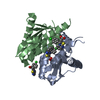
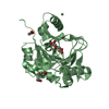
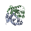
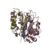
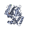
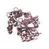
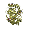
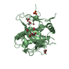
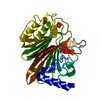
| Title | Core domain of HIV-1 integrase complexed with Mg++ and 1-(5-chloroindol-3-yl)-3-hydroxy-3-(2H-tetrazol-5-yl)-propenone | ||||||
 Components Components | PROTEIN (HIV-1 INTEGRASE (E.C.2.7.7.49)) | ||||||
 Keywords Keywords | TRANSFERASE / DNA INTEGRATION / INTEGRASE / HIV / ASPARTYL PROTEASE / ENDONUCLEASE | ||||||
| Function / homology |  Function and homology information Function and homology informationHIV-1 retropepsin / symbiont-mediated activation of host apoptosis / retroviral ribonuclease H / exoribonuclease H / exoribonuclease H activity / DNA integration / viral genome integration into host DNA / establishment of integrated proviral latency / RNA-directed DNA polymerase / RNA stem-loop binding ...HIV-1 retropepsin / symbiont-mediated activation of host apoptosis / retroviral ribonuclease H / exoribonuclease H / exoribonuclease H activity / DNA integration / viral genome integration into host DNA / establishment of integrated proviral latency / RNA-directed DNA polymerase / RNA stem-loop binding / viral penetration into host nucleus / host multivesicular body / RNA-directed DNA polymerase activity / RNA-DNA hybrid ribonuclease activity / Transferases; Transferring phosphorus-containing groups; Nucleotidyltransferases / host cell / viral nucleocapsid / endonuclease activity / DNA recombination / DNA-directed DNA polymerase / aspartic-type endopeptidase activity / Hydrolases; Acting on ester bonds / host cell cytoplasm / DNA-directed DNA polymerase activity / symbiont-mediated suppression of host gene expression / viral translational frameshifting / symbiont entry into host cell / lipid binding / host cell nucleus / host cell plasma membrane / virion membrane / structural molecule activity / proteolysis / DNA binding / zinc ion binding Similarity search - Function | ||||||
| Biological species |   Human immunodeficiency virus 1 Human immunodeficiency virus 1 | ||||||
| Method |  X-RAY DIFFRACTION / Resolution: 2.1 Å X-RAY DIFFRACTION / Resolution: 2.1 Å | ||||||
 Authors Authors | Goldgur, Y. / Craigie, R. / Fujiwara, T. / Yoshinaga, T. / Davies, D.R. | ||||||
 Citation Citation | Journal: Proc.Natl.Acad.Sci.USA / Year: 1999 Title: Structure of the HIV-1 integrase catalytic domain complexed with an inhibitor: a platform for antiviral drug design. Authors: Goldgur, Y. / Craigie, R. / Cohen, G.H. / Fujiwara, T. / Yoshinaga, T. / Fujishita, T. / Sugimoto, H. / Endo, T. / Murai, H. / Davies, D.R. | ||||||
| History |
|
- Structure visualization
| Structure viewer | Molecule: MolmilJmol/JSmol |
|---|
- Downloads & links
-Download
| PDBx/mmCIF format | 1qs4.cif.gz | 112.1 KB | Display | PDBx/mmCIF format |
|---|---|---|---|---|
| PDB format | pdb1qs4.ent.gz | 85.4 KB | Display | PDB format |
| PDBx/mmJSON format | 1qs4.json.gz | Tree view | PDBx/mmJSON format | |
| Others |  Other downloads Other downloads |
-Validation report
| Arichive directory | https://data.pdbj.org/pub/pdb/validation_reports/qs/1qs4ftp://data.pdbj.org/pub/pdb/validation_reports/qs/1qs4 | HTTPS FTP |
|---|
-Related structure data
| Similar structure data |
|---|

-Links
 PDBj
PDBj

- Assembly
| Deposited unit | 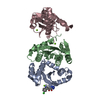
| ||||||||
|---|---|---|---|---|---|---|---|---|---|
| 1 | 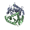
| ||||||||
| 2 | 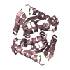
| ||||||||
| Unit cell |
|
-Components
| #1: Protein | Mass: 16813.219 Da / Num. of mol.: 3 / Fragment: Catalytic core domain / Mutation: F185K, W131E Source method: isolated from a genetically manipulated source Source: (gene. exp.) Human immunodeficiency virus 1 / Genus: Lentivirus / Plasmid: PET15B / Species (production host): Escherichia coli / Production host:  References: UniProt: Q76353, UniProt: P12497*PLUS, RNA-directed DNA polymerase #2: Chemical |   Mass: 24.305 Da / Num. of mol.: 3 / Source method: obtained synthetically / Formula: Mg Mass: 24.305 Da / Num. of mol.: 3 / Source method: obtained synthetically / Formula: Mg#3: Chemical | ChemComp-100 / |   Mass: 289.677 Da / Num. of mol.: 1 / Source method: obtained synthetically / Formula: C12H8ClN5O2 Mass: 289.677 Da / Num. of mol.: 1 / Source method: obtained synthetically / Formula: C12H8ClN5O2#4: Water | ChemComp-HOH / |  Mass: 18.015 Da / Num. of mol.: 544 / Source method: isolated from a natural source / Formula: H2O Mass: 18.015 Da / Num. of mol.: 544 / Source method: isolated from a natural source / Formula: H2O |
|---|
-Experimental details
-Experiment
| Experiment | Method: X-RAY DIFFRACTION / Number of used crystals: 1 |
|---|
- Sample preparation
| Crystal | Density Matthews: 2.4 Å3/Da / Density % sol: 48.84 % | ||||||||||||||||||||||||||||||||||||||||||||||||||||||||||||
|---|---|---|---|---|---|---|---|---|---|---|---|---|---|---|---|---|---|---|---|---|---|---|---|---|---|---|---|---|---|---|---|---|---|---|---|---|---|---|---|---|---|---|---|---|---|---|---|---|---|---|---|---|---|---|---|---|---|---|---|---|---|
| Crystal grow | pH: 7 Details: 30% PEG 4000, 100 MM HEPES, 5 MM MGCL2, 5 MM DTT, 1% GLYCEROL, pH 7.00 | ||||||||||||||||||||||||||||||||||||||||||||||||||||||||||||
| Crystal grow | *PLUS pH: 7.5 / Method: vapor diffusion, hanging dropDetails: Goldgur, Y., (1998) Proc. Natl. Acad. Sci. U.S.A., 95, 9150. | ||||||||||||||||||||||||||||||||||||||||||||||||||||||||||||
| Components of the solutions | *PLUS
|
-Data collection
| Diffraction | Mean temperature: 95 K |
|---|---|
| Diffraction source | Source: ROTATING ANODE / Type: RIGAKU RU200 / Wavelength: 1.5418 |
| Detector | Type: RIGAKU RAXIS IV / Detector: IMAGE PLATE / Date: Jan 1, 1999 |
| Radiation | Protocol: SINGLE WAVELENGTH / Monochromatic (M) / Laue (L): M / Scattering type: x-ray |
| Radiation wavelength | Wavelength: 1.5418 Å / Relative weight: 1 |
| Reflection | Resolution: 2.1→38.07 Å / Num. obs: 26564 / % possible obs: 94.2 % / Observed criterion σ(I): -3 / Redundancy: 2.12 % / Biso Wilson estimate: 29.64 Å2 / Rmerge(I) obs: 0.051 / Net I/σ(I): 10.4 |
| Reflection shell | Resolution: 2.1→2.18 Å / Redundancy: 1.5 % / Rmerge(I) obs: 0.274 / % possible all: 81.2 |
| Reflection | *PLUS Highest resolution: 2.1 Å / Lowest resolution: 20 Å / Observed criterion σ(I): -3 |
| Reflection shell | *PLUS % possible obs: 81 % / Mean I/σ(I) obs: 2.86 |
- Processing
| Software |
| ||||||||||||||||||||||||||||||||||||||||||||||||||||||||||||||||||||||||||||||||
|---|---|---|---|---|---|---|---|---|---|---|---|---|---|---|---|---|---|---|---|---|---|---|---|---|---|---|---|---|---|---|---|---|---|---|---|---|---|---|---|---|---|---|---|---|---|---|---|---|---|---|---|---|---|---|---|---|---|---|---|---|---|---|---|---|---|---|---|---|---|---|---|---|---|---|---|---|---|---|---|---|---|
| Refinement | Resolution: 2.1→38.07 Å / σ(F): 0 / Stereochemistry target values: Engh & Huber
| ||||||||||||||||||||||||||||||||||||||||||||||||||||||||||||||||||||||||||||||||
| Solvent computation | Solvent model: BABINET'S PRINCIPLE / Bsol: 73.9058 Å2 / ksol: 0.3627 e/Å3 | ||||||||||||||||||||||||||||||||||||||||||||||||||||||||||||||||||||||||||||||||
| Displacement parameters |
| ||||||||||||||||||||||||||||||||||||||||||||||||||||||||||||||||||||||||||||||||
| Refinement step | Cycle: LAST / Resolution: 2.1→38.07 Å
| ||||||||||||||||||||||||||||||||||||||||||||||||||||||||||||||||||||||||||||||||
| Refine LS restraints |
| ||||||||||||||||||||||||||||||||||||||||||||||||||||||||||||||||||||||||||||||||
| LS refinement shell | Resolution: 2.1→2.13 Å / Total num. of bins used: 25
| ||||||||||||||||||||||||||||||||||||||||||||||||||||||||||||||||||||||||||||||||
| Xplor file |
| ||||||||||||||||||||||||||||||||||||||||||||||||||||||||||||||||||||||||||||||||
| Software | *PLUS Name: CNS / Classification: refinement | ||||||||||||||||||||||||||||||||||||||||||||||||||||||||||||||||||||||||||||||||
| Refinement | *PLUS % reflection Rfree: 5 % | ||||||||||||||||||||||||||||||||||||||||||||||||||||||||||||||||||||||||||||||||
| Solvent computation | *PLUS | ||||||||||||||||||||||||||||||||||||||||||||||||||||||||||||||||||||||||||||||||
| Displacement parameters | *PLUS | ||||||||||||||||||||||||||||||||||||||||||||||||||||||||||||||||||||||||||||||||
| LS refinement shell | *PLUS Highest resolution: 2.1 Å / % reflection Rfree: 5 % |
