 Movie
Movie Controller

[English] 日本語
 Yorodumi
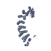
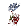
- PDB-2xt1: Crystal structure of the HIV-1 capsid protein C-terminal domain (... -
+ Open data

- Basic information
| Entry | Database: PDB / ID: 2xt1 | ||||||
|---|---|---|---|---|---|---|---|
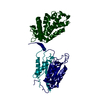
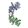
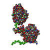
| Title | Crystal structure of the HIV-1 capsid protein C-terminal domain (146- 231) in complex with a camelid VHH. | ||||||
 Components Components |
| ||||||
 Keywords Keywords | VIRAL PROTEIN/IMMUNE SYSTEM / VIRAL PROTEIN-IMMUNE SYSTEM COMPLEX | ||||||
| Function / homology |  Function and homology information Function and homology informationviral nucleocapsid / host cell cytoplasm / viral translational frameshifting / host cell nucleus / host cell plasma membrane / RNA binding Similarity search - Function | ||||||
| Biological species |   HUMAN IMMUNODEFICIENCY VIRUS 1 HUMAN IMMUNODEFICIENCY VIRUS 1 | ||||||
| Method |  X-RAY DIFFRACTION / SYNCHROTRON / MOLECULAR REPLACEMENT / Resolution: 1.32 Å X-RAY DIFFRACTION / SYNCHROTRON / MOLECULAR REPLACEMENT / Resolution: 1.32 Å | ||||||
 Authors Authors | Igonet, S. / Vaney, M.C. / Bartonova, V. / Helma, J. / Rothbauer, U. / Leonhardt, H. / Stura, E. / Krausslich, H.-G. / Rey, F.A. | ||||||
 Citation Citation | Journal: To be Published Title: Targeting HIV-1 Virion Formation with Nanobodies -Implications for the Design of Assembly Inhibitors Authors: Igonet, S. / Vaney, M.C. / Bartonova, V. / Helma, J. / Rothbauer, U. / Leonhardt, H. / Stura, E. / Krausslich, H.-G. / Rey, F.A. | ||||||
| History |
|
- Structure visualization
| Structure viewer | Molecule: MolmilJmol/JSmol |
|---|
- Downloads & links
-Download
| PDBx/mmCIF format | 2xt1.cif.gz | 59.2 KB | Display | PDBx/mmCIF format |
|---|---|---|---|---|
| PDB format | pdb2xt1.ent.gz | 42.5 KB | Display | PDB format |
| PDBx/mmJSON format | 2xt1.json.gz | Tree view | PDBx/mmJSON format | |
| Others |  Other downloads Other downloads |
-Validation report
| Arichive directory | https://data.pdbj.org/pub/pdb/validation_reports/xt/2xt1ftp://data.pdbj.org/pub/pdb/validation_reports/xt/2xt1 | HTTPS FTP |
|---|
-Related structure data
| Related structure data |  2xv6C 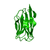 2xxcC  2xxmC 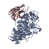 1kxvS  2buoS C: citing same article ( S: Starting model for refinement |
|---|---|
| Similar structure data |


-Links
 PDBj
PDBj


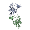
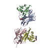
- Assembly
| Deposited unit | 
| |||||||||
|---|---|---|---|---|---|---|---|---|---|---|
| 1 | 
| |||||||||
| Unit cell |
| |||||||||
| Components on special symmetry positions |
|
-Components
| #1: Protein | Mass: 9530.921 Da / Num. of mol.: 1 / Fragment: C-TERMINAL DOMAIN, RESIDUES 146-231 Source method: isolated from a genetically manipulated source Source: (gene. exp.) HUMAN IMMUNODEFICIENCY VIRUS 1 / Strain: NL4-3 / Plasmid: PET11C / Production host:  | ||||||
|---|---|---|---|---|---|---|---|
| #2: Antibody | Mass: 13213.812 Da / Num. of mol.: 1 Source method: isolated from a genetically manipulated source Source: (gene. exp.) | ||||||
| #3: Chemical |   Mass: 92.094 Da / Num. of mol.: 2 / Source method: obtained synthetically / Formula: C3H8O3 Mass: 92.094 Da / Num. of mol.: 2 / Source method: obtained synthetically / Formula: C3H8O3#4: Water | ChemComp-HOH / |  Mass: 18.015 Da / Num. of mol.: 271 / Source method: isolated from a natural source / Formula: H2O Mass: 18.015 Da / Num. of mol.: 271 / Source method: isolated from a natural source / Formula: H2OHas protein modification | Y | Sequence details | THE VHH RESIDUES (CHAIN B) ARE NUMBERED ACCORDING TO THE KABAT NUMBERING. | |
-Experimental details
-Experiment
| Experiment | Method: X-RAY DIFFRACTION / Number of used crystals: 5 |
|---|
- Sample preparation
| Crystal | Density Matthews: 2.13 Å3/Da / Density % sol: 42.3 % / Description: NONE |
|---|---|
| Crystal grow | Details: 28-32 % W/V PEG 4000 |
-Data collection
| Diffraction | Mean temperature: 100 K |
|---|---|
| Diffraction source | Source: SYNCHROTRON / Site: ESRF  / Beamline: ID14-4 / Wavelength: 0.9395 / Beamline: ID14-4 / Wavelength: 0.9395 |
| Detector | Type: ADSC QUANTUM 315r / Detector: CCD / Date: Oct 15, 2008 |
| Radiation | Protocol: SINGLE WAVELENGTH / Monochromatic (M) / Laue (L): M / Scattering type: x-ray |
| Radiation wavelength | Wavelength: 0.9395 Å / Relative weight: 1 |
| Reflection | Resolution: 1.32→44.1 Å / Num. obs: 45032 / % possible obs: 98.2 % / Observed criterion σ(I): 2.1 / Redundancy: 13.7 % / Biso Wilson estimate: 10 Å2 / Rmerge(I) obs: 0.07 / Net I/σ(I): 14.7 |
| Reflection shell | Resolution: 1.32→1.39 Å / Redundancy: 5 % / Rmerge(I) obs: 0.46 / Mean I/σ(I) obs: 2.1 / % possible all: 88.6 |
- Processing
| Software |
| ||||||||||||||||||||||||||||||||||||||||||||||||||||||||||||||||||||||||||||||||||||||||||||||||||||||||||||||||||
|---|---|---|---|---|---|---|---|---|---|---|---|---|---|---|---|---|---|---|---|---|---|---|---|---|---|---|---|---|---|---|---|---|---|---|---|---|---|---|---|---|---|---|---|---|---|---|---|---|---|---|---|---|---|---|---|---|---|---|---|---|---|---|---|---|---|---|---|---|---|---|---|---|---|---|---|---|---|---|---|---|---|---|---|---|---|---|---|---|---|---|---|---|---|---|---|---|---|---|---|---|---|---|---|---|---|---|---|---|---|---|---|---|---|---|---|
| Refinement | Method to determine structure: MOLECULAR REPLACEMENT Starting model: PDB ENTRIES 2BUO, 1KXV Resolution: 1.32→35.54 Å / Cor.coef. Fo:Fc: 0.9647 / Cor.coef. Fo:Fc free: 0.9558 / SU R Cruickshank DPI: 0.048 / Cross valid method: THROUGHOUT / σ(F): 0 / SU R Blow DPI: 0.052 / SU Rfree Blow DPI: 0.053 / SU Rfree Cruickshank DPI: 0.05 Details: THE 6XHISTIDINE TAG AT THE C-TERMINAL END OF CHAIN B IS DISORDERED.
| ||||||||||||||||||||||||||||||||||||||||||||||||||||||||||||||||||||||||||||||||||||||||||||||||||||||||||||||||||
| Displacement parameters | Biso mean: 15.57 Å2
| ||||||||||||||||||||||||||||||||||||||||||||||||||||||||||||||||||||||||||||||||||||||||||||||||||||||||||||||||||
| Refine analyze | Luzzati coordinate error obs: 0.129 Å | ||||||||||||||||||||||||||||||||||||||||||||||||||||||||||||||||||||||||||||||||||||||||||||||||||||||||||||||||||
| Refinement step | Cycle: LAST / Resolution: 1.32→35.54 Å
| ||||||||||||||||||||||||||||||||||||||||||||||||||||||||||||||||||||||||||||||||||||||||||||||||||||||||||||||||||
| Refine LS restraints |
| ||||||||||||||||||||||||||||||||||||||||||||||||||||||||||||||||||||||||||||||||||||||||||||||||||||||||||||||||||
| LS refinement shell | Resolution: 1.32→1.35 Å / Total num. of bins used: 20
|
