 Movie
Movie Controller


+ Open data

- Basic information
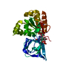
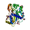
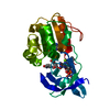
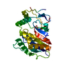
| Entry | Database: PDB / ID: 2atp | ||||||
|---|---|---|---|---|---|---|---|
| Title | Crystal structure of a CD8ab heterodimer | ||||||
 Components Components |
| ||||||
 Keywords Keywords | IMMUNE SYSTEM / CD8ab / CD8aa / MHC | ||||||
| Function / homology |  Function and homology information Function and homology informationcytotoxic T cell differentiation / MHC class I protein complex binding / T cell mediated immunity / Immunoregulatory interactions between a Lymphoid and a non-Lymphoid cell / MHC class I protein binding / plasma membrane raft / regulation of immune response / coreceptor activity / positive regulation of calcium-mediated signaling / T cell activation ...cytotoxic T cell differentiation / MHC class I protein complex binding / T cell mediated immunity / Immunoregulatory interactions between a Lymphoid and a non-Lymphoid cell / MHC class I protein binding / plasma membrane raft / regulation of immune response / coreceptor activity / positive regulation of calcium-mediated signaling / T cell activation / T cell receptor signaling pathway / defense response to virus / adaptive immune response / cell surface receptor signaling pathway / signaling receptor complex / external side of plasma membrane / protein kinase binding / cell surface / identical protein binding / plasma membrane Similarity search - Function | ||||||
| Biological species |  | ||||||
| Method |  X-RAY DIFFRACTION / SYNCHROTRON / MOLECULAR REPLACEMENT / Resolution: 2.4 Å X-RAY DIFFRACTION / SYNCHROTRON / MOLECULAR REPLACEMENT / Resolution: 2.4 Å | ||||||
 Authors Authors | Chang, H.C. / Tan, K. / Ouyang, J. / Parisini, E. / Liu, J.H. / Le, Y. / Wang, X. / Reinherz, E.L. / Wang, J.H. | ||||||
 Citation Citation | Journal: Immunity / Year: 2005 Title: Structural and Mutational Analyses of a CD8alphabeta Heterodimer and Comparison with the CD8alphaalpha Homodimer. Authors: Chang, H.C. / Tan, K. / Ouyang, J. / Parisini, E. / Liu, J.H. / Le, Y. / Wang, X. / Reinherz, E.L. / Wang, J.H. | ||||||
| History |
|
- Structure visualization
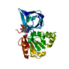
| Structure viewer | Molecule: MolmilJmol/JSmol |
|---|
- Downloads & links
-Download
| PDBx/mmCIF format | 2atp.cif.gz | 108.8 KB | Display | PDBx/mmCIF format |
|---|---|---|---|---|
| PDB format | pdb2atp.ent.gz | 84 KB | Display | PDB format |
| PDBx/mmJSON format | 2atp.json.gz | Tree view | PDBx/mmJSON format | |
| Others |  Other downloads Other downloads |
-Validation report
| Arichive directory | https://data.pdbj.org/pub/pdb/validation_reports/at/2atpftp://data.pdbj.org/pub/pdb/validation_reports/at/2atp | HTTPS FTP |
|---|
-Related structure data
| Related structure data |  1bqhS S: Starting model for refinement |
|---|---|
| Similar structure data |


-Links
 PDBj
PDBj


- Assembly
| Deposited unit | 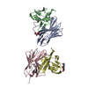
| ||||||||
|---|---|---|---|---|---|---|---|---|---|
| 1 | 
| ||||||||
| 2 | 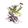
| ||||||||
| Unit cell |
|
-Components
| #1: Protein | Mass: 13842.911 Da / Num. of mol.: 2 / Fragment: CD8a ectodomain fragment Source method: isolated from a genetically manipulated source Source: (gene. exp.)  Cricetulus griseus (Chinese hamster) / Strain (production host): CHO-Lec 3.8.2.1 / References: UniProt: P01731 Cricetulus griseus (Chinese hamster) / Strain (production host): CHO-Lec 3.8.2.1 / References: UniProt: P01731#2: Protein/peptide | Mass: 3028.172 Da / Num. of mol.: 2 Fragment: a peptide used to connect CD8a C-terminal and CD8b N-terminal Source method: obtained synthetically / Details: synthetic linker #3: Protein | Mass: 13038.967 Da / Num. of mol.: 2 / Fragment: CD8b ectodomain fragment Source method: isolated from a genetically manipulated source Source: (gene. exp.) Cricetulus griseus (Chinese hamster) / Strain (production host): CHO-Lec 3.8.2.1 / References: UniProt: P10300#4: Sugar |   Type: D-saccharide, beta linking / Mass: 221.208 Da / Num. of mol.: 3 Type: D-saccharide, beta linking / Mass: 221.208 Da / Num. of mol.: 3Source method: isolated from a genetically manipulated source Formula: C8H15NO6 #5: Water | ChemComp-HOH / |  Mass: 18.015 Da / Num. of mol.: 44 / Source method: isolated from a natural source / Formula: H2O Mass: 18.015 Da / Num. of mol.: 44 / Source method: isolated from a natural source / Formula: H2OHas protein modification | Y | |
|---|
-Experimental details
-Experiment
| Experiment | Method: X-RAY DIFFRACTION / Number of used crystals: 1 |
|---|
- Sample preparation
| Crystal | Density Matthews: 2.3 Å3/Da / Density % sol: 46.2 % |
|---|---|
| Crystal grow | Temperature: 293 K / Method: vapor diffusion, hanging drop / pH: 8.5 Details: 25% PEG4000, 0.2M (NH4)2SO4, 0.1 M Tris , pH 8.5, VAPOR DIFFUSION, HANGING DROP, temperature 293K |
-Data collection
| Diffraction | Mean temperature: 200 K |
|---|---|
| Diffraction source | Source: SYNCHROTRON / Site: APS  / Beamline: 19-ID / Wavelength: 0.9791 Å / Beamline: 19-ID / Wavelength: 0.9791 Å |
| Detector | Type: ADSC QUANTUM 315 / Detector: CCD / Date: Dec 5, 2004 |
| Radiation | Monochromator: graphite / Protocol: SINGLE WAVELENGTH / Monochromatic (M) / Laue (L): M / Scattering type: x-ray |
| Radiation wavelength | Wavelength: 0.9791 Å / Relative weight: 1 |
| Reflection | Resolution: 2.4→50 Å / Num. all: 21001 / Num. obs: 21001 / % possible obs: 99.3 % / Observed criterion σ(F): 0 / Observed criterion σ(I): 0 / Redundancy: 3.5 % / Biso Wilson estimate: 51.2 Å2 / Rmerge(I) obs: 0.07 / Net I/σ(I): 17.8 |
| Reflection shell | Resolution: 2.4→2.49 Å / Redundancy: 3.1 % / Rmerge(I) obs: 0.44 / Mean I/σ(I) obs: 2.1 / Num. unique all: 2071 / % possible all: 96.8 |
- Processing
| Software |
| |||||||||||||||||||||||||
|---|---|---|---|---|---|---|---|---|---|---|---|---|---|---|---|---|---|---|---|---|---|---|---|---|---|---|
| Refinement | Method to determine structure: MOLECULAR REPLACEMENT Starting model: PDB ENTRY 1BQH Resolution: 2.4→25 Å / Isotropic thermal model: Isotropic / σ(F): 1 / Stereochemistry target values: Engh & Huber
| |||||||||||||||||||||||||
| Displacement parameters | Biso mean: 52.314 Å2 | |||||||||||||||||||||||||
| Refine analyze | Luzzati coordinate error obs: 0.38 Å / Luzzati d res low obs: 5 Å / Luzzati sigma a obs: 0.6 Å | |||||||||||||||||||||||||
| Refinement step | Cycle: LAST / Resolution: 2.4→25 Å
| |||||||||||||||||||||||||
| Refine LS restraints |
| |||||||||||||||||||||||||
| LS refinement shell | Resolution: 2.4→2.49 Å
|
