 Movie
Movie Controller


+ Open data

- Basic information
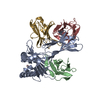
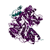
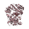
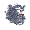
| Entry | Database: PDB / ID: 1bqh | |||||||||
|---|---|---|---|---|---|---|---|---|---|---|
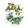
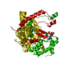
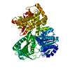
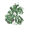
| Title | MURINE CD8AA ECTODOMAIN FRAGMENT IN COMPLEX WITH H-2KB/VSV8 | |||||||||
 Components Components |
| |||||||||
 Keywords Keywords | IMMUNE SYSTEM / T-CELL / CORECEPTOR / GLYCOPROTEIN / COMPLEX / IMMUNOLOGY / ANTIGEN | |||||||||
| Function / homology |  Function and homology information Function and homology informationcytotoxic T cell differentiation / MHC class I protein complex binding / T cell mediated immunity / MHC class Ib protein complex / natural killer cell lectin-like receptor binding / TAP2 binding / TAP1 binding / cis-Golgi network membrane / Endosomal/Vacuolar pathway / DAP12 interactions ...cytotoxic T cell differentiation / MHC class I protein complex binding / T cell mediated immunity / MHC class Ib protein complex / natural killer cell lectin-like receptor binding / TAP2 binding / TAP1 binding / cis-Golgi network membrane / Endosomal/Vacuolar pathway / DAP12 interactions / Antigen Presentation: Folding, assembly and peptide loading of class I MHC / ER-Phagosome pathway / DAP12 signaling / Immunoregulatory interactions between a Lymphoid and a non-Lymphoid cell / antigen processing and presentation of endogenous peptide antigen via MHC class I via ER pathway, TAP-dependent / TAP complex binding / antigen processing and presentation of exogenous peptide antigen via MHC class I / Golgi medial cisterna / regulation of membrane depolarization / inner ear development / CD8 receptor binding / endoplasmic reticulum exit site / TAP binding / beta-2-microglobulin binding / MHC class I protein binding / plasma membrane raft / antigen processing and presentation of endogenous peptide antigen via MHC class Ib / antigen processing and presentation of endogenous peptide antigen via MHC class I via ER pathway, TAP-independent / cellular defense response / T cell receptor binding / positive regulation of calcium-mediated signaling / Neutrophil degranulation / 14-3-3 protein binding / T cell activation / lumenal side of endoplasmic reticulum membrane / regulation of iron ion transport / cellular response to iron(III) ion / antigen processing and presentation of exogenous protein antigen via MHC class Ib, TAP-dependent / negative regulation of iron ion transport / negative regulation of forebrain neuron differentiation / regulation of erythrocyte differentiation / iron ion transport / peptide antigen assembly with MHC class I protein complex / response to molecule of bacterial origin / HFE-transferrin receptor complex / MHC class I peptide loading complex / transferrin transport / negative regulation of receptor-mediated endocytosis / cellular response to iron ion / positive regulation of T cell cytokine production / antigen processing and presentation of endogenous peptide antigen via MHC class I / MHC class I protein complex / peptide antigen assembly with MHC class II protein complex / negative regulation of neurogenesis / multicellular organismal-level iron ion homeostasis / cellular response to nicotine / MHC class II protein complex / positive regulation of receptor-mediated endocytosis / positive regulation of T cell mediated cytotoxicity / negative regulation of epithelial cell proliferation / antigen processing and presentation of exogenous peptide antigen via MHC class II / positive regulation of immune response / peptide antigen binding / phagocytic vesicle membrane / positive regulation of T cell activation / sensory perception of smell / positive regulation of cellular senescence / MHC class II protein complex binding / T cell differentiation in thymus / T cell receptor signaling pathway / late endosome membrane / negative regulation of neuron projection development / antimicrobial humoral immune response mediated by antimicrobial peptide / antibacterial humoral response / cellular response to lipopolysaccharide / protein refolding / protein-folding chaperone binding / early endosome membrane / defense response to virus / amyloid fibril formation / defense response to Gram-negative bacterium / protein homotetramerization / intracellular iron ion homeostasis / adaptive immune response / learning or memory / early endosome / cell surface receptor signaling pathway / signaling receptor complex / defense response to bacterium / defense response to Gram-positive bacterium / immune response / receptor ligand activity / Golgi membrane / external side of plasma membrane / signaling receptor binding / innate immune response / lysosomal membrane / protein kinase binding / structural molecule activity / Golgi apparatus Similarity search - Function | |||||||||
| Biological species |  | |||||||||
| Method |  X-RAY DIFFRACTION / SYNCHROTRON / MOLECULAR REPLACEMENT / Resolution: 2.8 Å X-RAY DIFFRACTION / SYNCHROTRON / MOLECULAR REPLACEMENT / Resolution: 2.8 Å | |||||||||
 Authors Authors | Wang, J.H. / Reinherz, E.L. / Kern, P.S. / Chang, H.C. | |||||||||
 Citation Citation | Journal: Immunity / Year: 1998 Title: Structural basis of CD8 coreceptor function revealed by crystallographic analysis of a murine CD8alphaalpha ectodomain fragment in complex with H-2Kb. Authors: Kern, P.S. / Teng, M.K. / Smolyar, A. / Liu, J.H. / Liu, J. / Hussey, R.E. / Spoerl, R. / Chang, H.C. / Reinherz, E.L. / Wang, J.H. | |||||||||
| History |
|
- Structure visualization
| Structure viewer | Molecule: MolmilJmol/JSmol |
|---|
- Downloads & links
-Download
| PDBx/mmCIF format | 1bqh.cif.gz | 259.5 KB | Display | PDBx/mmCIF format |
|---|---|---|---|---|
| PDB format | pdb1bqh.ent.gz | 209.6 KB | Display | PDB format |
| PDBx/mmJSON format | 1bqh.json.gz | Tree view | PDBx/mmJSON format | |
| Others |  Other downloads Other downloads |
-Validation report
| Arichive directory | https://data.pdbj.org/pub/pdb/validation_reports/bq/1bqhftp://data.pdbj.org/pub/pdb/validation_reports/bq/1bqh | HTTPS FTP |
|---|
-Related structure data
| Related structure data |  2vaaS S: Starting model for refinement |
|---|---|
| Similar structure data |


-Links
 PDBj
PDBj


- Assembly
| Deposited unit | 
| ||||||||
|---|---|---|---|---|---|---|---|---|---|
| 1 | 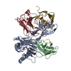
| ||||||||
| 2 | 
| ||||||||
| Unit cell |
|
-Components
| #1: Protein | Mass: 31648.322 Da / Num. of mol.: 2 / Fragment: ALPHA CHAIN / Source method: isolated from a natural source / Source: (natural) #2: Protein | Mass: 11704.359 Da / Num. of mol.: 2 / Fragment: BETA CHAIN / Source method: isolated from a natural source / Source: (natural) #3: Protein/peptide | Mass: 956.078 Da / Num. of mol.: 2 / Fragment: ANTIGENIC PEPTIDE / Source method: obtained synthetically #4: Protein | Mass: 14512.637 Da / Num. of mol.: 4 / Fragment: IG ECTODOMAIN FRAGMENT Source method: isolated from a genetically manipulated source Source: (gene. exp.) Description: GENE FUSED TO THROMBIN CLEAVAGE SITE AND LEUCINE ZIPPER Cell line (production host): CHO LEC 3.2.8.1 / Gene (production host): CD8A / Production host: #5: Sugar | ChemComp-NAG /   Type: D-saccharide, beta linking / Mass: 221.208 Da / Num. of mol.: 4 Type: D-saccharide, beta linking / Mass: 221.208 Da / Num. of mol.: 4Source method: isolated from a genetically manipulated source Formula: C8H15NO6 Has protein modification | Y | |
|---|
-Experimental details
-Experiment
| Experiment | Method: X-RAY DIFFRACTION / Number of used crystals: 1 |
|---|
- Sample preparation
| Crystal | Density Matthews: 2.5 Å3/Da / Density % sol: 51 % | ||||||||||||||||||||||||||||||||||||
|---|---|---|---|---|---|---|---|---|---|---|---|---|---|---|---|---|---|---|---|---|---|---|---|---|---|---|---|---|---|---|---|---|---|---|---|---|---|
| Crystal grow | pH: 8.5 / Details: 20-25% PEG 4K, 0.2M LI2SO4, 0.1M TRIS PH8.5 | ||||||||||||||||||||||||||||||||||||
| Crystal grow | *PLUS pH: 7 / Method: vapor diffusion, hanging drop | ||||||||||||||||||||||||||||||||||||
| Components of the solutions | *PLUS
|
-Data collection
| Diffraction | Mean temperature: 110 K |
|---|---|
| Diffraction source | Source: SYNCHROTRON / Site: NSLS  / Beamline: X12C / Wavelength: 1.15 / Beamline: X12C / Wavelength: 1.15 |
| Detector | Type: BRANDEIS / Detector: CCD / Date: Apr 15, 1997 |
| Radiation | Protocol: SINGLE WAVELENGTH / Monochromatic (M) / Laue (L): M / Scattering type: x-ray |
| Radiation wavelength | Wavelength: 1.15 Å / Relative weight: 1 |
| Reflection | Resolution: 2.8→15 Å / Num. obs: 34131 / % possible obs: 93.8 % / Observed criterion σ(I): 2 / Redundancy: 2.9 % / Rsym value: 0.061 / Net I/σ(I): 9.7 |
| Reflection shell | Resolution: 2.8→2.9 Å / Mean I/σ(I) obs: 1.9 / Rsym value: 0.249 / % possible all: 90.7 |
| Reflection | *PLUS Num. measured all: 99259 / Rmerge(I) obs: 0.061 |
| Reflection shell | *PLUS % possible obs: 90.7 % / Rmerge(I) obs: 0.249 |
- Processing
| Software |
| ||||||||||||||||||||||||||||||||||||||||||||||||||||||||||||
|---|---|---|---|---|---|---|---|---|---|---|---|---|---|---|---|---|---|---|---|---|---|---|---|---|---|---|---|---|---|---|---|---|---|---|---|---|---|---|---|---|---|---|---|---|---|---|---|---|---|---|---|---|---|---|---|---|---|---|---|---|---|
| Refinement | Method to determine structure: MOLECULAR REPLACEMENT Starting model: PDB ENTRY 2VAA Resolution: 2.8→15 Å / Cross valid method: THROUGHOUT / σ(F): 2
| ||||||||||||||||||||||||||||||||||||||||||||||||||||||||||||
| Refinement step | Cycle: LAST / Resolution: 2.8→15 Å
| ||||||||||||||||||||||||||||||||||||||||||||||||||||||||||||
| Refine LS restraints |
| ||||||||||||||||||||||||||||||||||||||||||||||||||||||||||||
| Refine LS restraints NCS | NCS model details: RESTRAINTS | ||||||||||||||||||||||||||||||||||||||||||||||||||||||||||||
| Xplor file |
| ||||||||||||||||||||||||||||||||||||||||||||||||||||||||||||
| Software | *PLUS Name: X-PLOR / Version: 3.8 / Classification: refinement | ||||||||||||||||||||||||||||||||||||||||||||||||||||||||||||
| Refinement | *PLUS Highest resolution: 2.8 Å / Lowest resolution: 15 Å / σ(F): 2 / % reflection Rfree: 10.1 % | ||||||||||||||||||||||||||||||||||||||||||||||||||||||||||||
| Solvent computation | *PLUS | ||||||||||||||||||||||||||||||||||||||||||||||||||||||||||||
| Displacement parameters | *PLUS | ||||||||||||||||||||||||||||||||||||||||||||||||||||||||||||
| Refine LS restraints | *PLUS
|
