 Movie
Movie Controller

[English] 日本語
 Yorodumi
- PDB-3ezn: Crystal structure of phosphoglyceromutase from burkholderia pseud... -
+ Open data

- Basic information
| Entry | Database: PDB / ID: 3ezn | ||||||
|---|---|---|---|---|---|---|---|
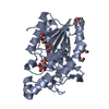
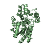
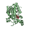
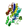
| Title | Crystal structure of phosphoglyceromutase from burkholderia pseudomallei 1710b | ||||||
 Components Components | 2,3-bisphosphoglycerate-dependent phosphoglycerate mutase | ||||||
 Keywords Keywords | ISOMERASE / SSGCID / PHOSPHOGLYCEROMUTASE / BURKHOLDERIA PSEUDOMALLEI / Glycolysis / Structural Genomics / Seattle Structural Genomics Center for Infectious Disease | ||||||
| Function / homology |  Function and homology information Function and homology informationphosphoglycerate mutase (2,3-diphosphoglycerate-dependent) / phosphoglycerate mutase activity / glycolytic process / gluconeogenesis Similarity search - Function | ||||||
| Biological species |  Burkholderia pseudomallei 1710b (bacteria) Burkholderia pseudomallei 1710b (bacteria) | ||||||
| Method |  X-RAY DIFFRACTION / MOLECULAR REPLACEMENT / Resolution: 2.1 Å X-RAY DIFFRACTION / MOLECULAR REPLACEMENT / Resolution: 2.1 Å | ||||||
 Authors Authors | Seattle Structural Genomics Center for Infectious Disease (SSGCID) | ||||||
 Citation Citation | Journal: Acta Crystallogr.,Sect.F / Year: 2011 Title: An ensemble of structures of Burkholderia pseudomallei 2,3-bisphosphoglycerate-dependent phosphoglycerate mutase. Authors: Davies, D.R. / Staker, B.L. / Abendroth, J.A. / Edwards, T.E. / Hartley, R. / Leonard, J. / Kim, H. / Rychel, A.L. / Hewitt, S.N. / Myler, P.J. / Stewart, L.J. | ||||||
| History |
|
- Structure visualization
| Structure viewer | Molecule: MolmilJmol/JSmol |
|---|
- Downloads & links
-Download
| PDBx/mmCIF format | 3ezn.cif.gz | 115.4 KB | Display | PDBx/mmCIF format |
|---|---|---|---|---|
| PDB format | pdb3ezn.ent.gz | 88.5 KB | Display | PDB format |
| PDBx/mmJSON format | 3ezn.json.gz | Tree view | PDBx/mmJSON format | |
| Others |  Other downloads Other downloads |
-Validation report
| Arichive directory | https://data.pdbj.org/pub/pdb/validation_reports/ez/3eznftp://data.pdbj.org/pub/pdb/validation_reports/ez/3ezn | HTTPS FTP |
|---|
-Related structure data
| Related structure data |  3fdzC  3gp3C  3gp5C  3gw8C  3lntC 1x19S C: citing same article ( S: Starting model for refinement |
|---|---|
| Similar structure data | |
| Other databases |

-Links
 PDBj
PDBj


- Assembly
| Deposited unit | 
| ||||||||||||||||||
|---|---|---|---|---|---|---|---|---|---|---|---|---|---|---|---|---|---|---|---|
| 1 | 
| ||||||||||||||||||
| 2 | 
| ||||||||||||||||||
| 3 |
| ||||||||||||||||||
| Unit cell |
| ||||||||||||||||||
| Noncrystallographic symmetry (NCS) | NCS domain:
NCS domain segments: Component-ID: 1 / Ens-ID: 1 / Beg auth comp-ID: MET / Beg label comp-ID: MET / End auth comp-ID: PG4 / End label comp-ID: PG4 / Refine code: 6 / Auth seq-ID: 1 - 250 / Label seq-ID: 9
NCS ensembles : (Details: A B) |
-Components
| #1: Protein | Mass: 28958.844 Da / Num. of mol.: 2 Source method: isolated from a genetically manipulated source Source: (gene. exp.) Burkholderia pseudomallei 1710b (bacteria)Strain: 1719B / Gene: gpmA, BURPS1710b_0662 / Plasmid: AVA0421 / Production host: #2: Chemical | ChemComp-PG4 /   Mass: 194.226 Da / Num. of mol.: 4 / Source method: obtained synthetically / Formula: C8H18O5 / Comment: precipitant*YM Mass: 194.226 Da / Num. of mol.: 4 / Source method: obtained synthetically / Formula: C8H18O5 / Comment: precipitant*YM#3: Water | ChemComp-HOH / |  Mass: 18.015 Da / Num. of mol.: 365 / Source method: isolated from a natural source / Formula: H2O Mass: 18.015 Da / Num. of mol.: 365 / Source method: isolated from a natural source / Formula: H2O |
|---|
-Experimental details
-Experiment
| Experiment | Method: X-RAY DIFFRACTION / Number of used crystals: 1 |
|---|
- Sample preparation
| Crystal | Density Matthews: 2.06 Å3/Da / Density % sol: 40.37 % |
|---|---|
| Crystal grow | Temperature: 290 K / Method: vapor diffusion, sitting drop / pH: 7.5 Details: EMERALD CRYO B-4: 100MM MES PH 6.0, 5% PEG 1000, 10% GLYCEROL, 30% PEG 600, PH 7.5, VAPOR DIFFUSION, TEMPERATURE 298K, pH 7.50, VAPOR DIFFUSION, SITTING DROP, temperature 290K |
-Data collection
| Diffraction | Mean temperature: 100 K |
|---|---|
| Diffraction source | Source: ROTATING ANODE / Type: RIGAKU MICROMAX-007 HF / Wavelength: 1.5418 |
| Detector | Type: SATURN 944 / Detector: CCD / Date: Sep 26, 2008 |
| Radiation | Protocol: SINGLE WAVELENGTH / Monochromatic (M) / Laue (L): M / Scattering type: x-ray |
| Radiation wavelength | Wavelength: 1.5418 Å / Relative weight: 1 |
| Reflection | Resolution: 2.1→50 Å / Num. all: 26261 / Num. obs: 26261 / % possible obs: 93.7 % / Observed criterion σ(F): 0 / Observed criterion σ(I): -3 / Redundancy: 3.74 % / Biso Wilson estimate: 22.93 Å2 / Rmerge(I) obs: 0.041 / Rsym value: 0.041 / Net I/σ(I): 25.7 |
| Reflection shell | Resolution: 2.1→2.15 Å / Redundancy: 3.6 % / Rmerge(I) obs: 0.137 / Mean I/σ(I) obs: 9.9 / Num. unique all: 1736 / Rsym value: 0.137 / % possible all: 83.2 |
- Processing
| Software |
| |||||||||||||||||||||||||||||||||||||||||||||||||||||||||||||||||||||||||||||||||||||
|---|---|---|---|---|---|---|---|---|---|---|---|---|---|---|---|---|---|---|---|---|---|---|---|---|---|---|---|---|---|---|---|---|---|---|---|---|---|---|---|---|---|---|---|---|---|---|---|---|---|---|---|---|---|---|---|---|---|---|---|---|---|---|---|---|---|---|---|---|---|---|---|---|---|---|---|---|---|---|---|---|---|---|---|---|---|---|
| Refinement | Method to determine structure: MOLECULAR REPLACEMENT Starting model: 1x19 modified with ccp4 chainsaw Resolution: 2.1→50 Å / Cor.coef. Fo:Fc: 0.957 / Cor.coef. Fo:Fc free: 0.925 / SU B: 4.047 / SU ML: 0.11 / Isotropic thermal model: isotrpoic / Cross valid method: THROUGHOUT / σ(F): 0 / σ(I): 0 / ESU R: 0.228 / ESU R Free: 0.181 / Stereochemistry target values: MAXIMUM LIKELIHOOD / Details: HYDROGENS HAVE BEEN ADDED IN THE RIDING POSITIONS
| |||||||||||||||||||||||||||||||||||||||||||||||||||||||||||||||||||||||||||||||||||||
| Solvent computation | Ion probe radii: 0.8 Å / Shrinkage radii: 0.8 Å / VDW probe radii: 1.2 Å / Solvent model: MASK | |||||||||||||||||||||||||||||||||||||||||||||||||||||||||||||||||||||||||||||||||||||
| Displacement parameters | Biso mean: 14.88 Å2
| |||||||||||||||||||||||||||||||||||||||||||||||||||||||||||||||||||||||||||||||||||||
| Refinement step | Cycle: LAST / Resolution: 2.1→50 Å
| |||||||||||||||||||||||||||||||||||||||||||||||||||||||||||||||||||||||||||||||||||||
| Refine LS restraints |
| |||||||||||||||||||||||||||||||||||||||||||||||||||||||||||||||||||||||||||||||||||||
| Refine LS restraints NCS | Dom-ID: 1 / Auth asym-ID: A / Ens-ID: 1 / Number: 3028 / Refine-ID: X-RAY DIFFRACTION
| |||||||||||||||||||||||||||||||||||||||||||||||||||||||||||||||||||||||||||||||||||||
| LS refinement shell | Resolution: 2.1→2.15 Å / Total num. of bins used: 20
|
