 Movie
Movie Controller

[English] 日本語
 Yorodumi
- PDB-3gp3: Crystal structure of phosphoglyceromutase from Burkholderia pseud... -
+ Open data

- Basic information
| Entry | Database: PDB / ID: 3gp3 | ||||||
|---|---|---|---|---|---|---|---|
| Title | Crystal structure of phosphoglyceromutase from Burkholderia pseudomallei with 2-phosphoserine | ||||||
 Components Components | 2,3-bisphosphoglycerate-dependent phosphoglycerate mutase | ||||||
 Keywords Keywords | ISOMERASE / phosphoglyceromutase / deCODE / SBRI / NIAID / UWPPG / Glycolysis / Structural Genomics / Seattle Structural Genomics Center for Infectious Disease / SSGCID | ||||||
| Function / homology |  Function and homology information Function and homology informationphosphoglycerate mutase (2,3-diphosphoglycerate-dependent) / phosphoglycerate mutase activity / glycolytic process / gluconeogenesis Similarity search - Function | ||||||
| Biological species |  Burkholderia pseudomallei (bacteria) Burkholderia pseudomallei (bacteria) | ||||||
| Method |  X-RAY DIFFRACTION / SYNCHROTRON / MOLECULAR REPLACEMENT / molecular replacement / Resolution: 1.5 Å X-RAY DIFFRACTION / SYNCHROTRON / MOLECULAR REPLACEMENT / molecular replacement / Resolution: 1.5 Å | ||||||
 Authors Authors | Seattle Structural Genomics Center for Infectious Disease (SSGCID) | ||||||
 Citation Citation | Journal: Acta Crystallogr.,Sect.F / Year: 2011 Title: An ensemble of structures of Burkholderia pseudomallei 2,3-bisphosphoglycerate-dependent phosphoglycerate mutase. Authors: Davies, D.R. / Staker, B.L. / Abendroth, J.A. / Edwards, T.E. / Hartley, R. / Leonard, J. / Kim, H. / Rychel, A.L. / Hewitt, S.N. / Myler, P.J. / Stewart, L.J. | ||||||
| History |
|
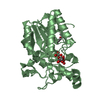
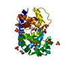
- Structure visualization
| Structure viewer | Molecule: MolmilJmol/JSmol |
|---|
- Downloads & links
-Download
| PDBx/mmCIF format | 3gp3.cif.gz | 212.4 KB | Display | PDBx/mmCIF format |
|---|---|---|---|---|
| PDB format | pdb3gp3.ent.gz | 169.9 KB | Display | PDB format |
| PDBx/mmJSON format | 3gp3.json.gz | Tree view | PDBx/mmJSON format | |
| Others |  Other downloads Other downloads |
-Validation report
| Arichive directory | https://data.pdbj.org/pub/pdb/validation_reports/gp/3gp3ftp://data.pdbj.org/pub/pdb/validation_reports/gp/3gp3 | HTTPS FTP |
|---|
-Related structure data
| Related structure data |  3eznC 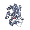 3fdzC  3gp5C  3gw8C  3lntC C: citing same article ( |
|---|---|
| Similar structure data | |
| Other databases |


-Links
 PDBj
PDBj


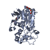
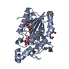
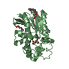
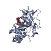
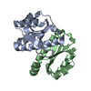
- Assembly
| Deposited unit | 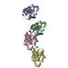
| ||||||||
|---|---|---|---|---|---|---|---|---|---|
| 1 | 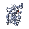
| ||||||||
| 2 | 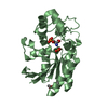
| ||||||||
| 3 | 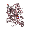
| ||||||||
| 4 | 
| ||||||||
| Unit cell |
|
-Components
| #1: Protein | Mass: 28958.844 Da / Num. of mol.: 4 Source method: isolated from a genetically manipulated source Details: Expressed with a non-cleavable N-terminal hexahis-tag Source: (gene. exp.) Burkholderia pseudomallei (bacteria) / Strain: 1710b / Gene: gpmA, BPSL0443 / Plasmid: BG1861 / Production host: References: UniProt: Q63XU7, UniProt: Q3JWH7*PLUS, EC: 5.4.2.1 #2: Chemical | ChemComp-PG4 /   Mass: 194.226 Da / Num. of mol.: 6 / Source method: obtained synthetically / Formula: C8H18O5 / Comment: precipitant*YM Mass: 194.226 Da / Num. of mol.: 6 / Source method: obtained synthetically / Formula: C8H18O5 / Comment: precipitant*YM#3: Chemical | ChemComp-PO3 /   Mass: 78.972 Da / Num. of mol.: 4 / Source method: obtained synthetically / Formula: PO3 Mass: 78.972 Da / Num. of mol.: 4 / Source method: obtained synthetically / Formula: PO3#4: Chemical | ChemComp-SEP / 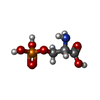  Type: L-peptide linking / Mass: 185.072 Da / Num. of mol.: 4 / Source method: obtained synthetically / Formula: C3H8NO6P Type: L-peptide linking / Mass: 185.072 Da / Num. of mol.: 4 / Source method: obtained synthetically / Formula: C3H8NO6P#5: Water | ChemComp-HOH / |  Mass: 18.015 Da / Num. of mol.: 712 / Source method: isolated from a natural source / Formula: H2O Mass: 18.015 Da / Num. of mol.: 712 / Source method: isolated from a natural source / Formula: H2O |
|---|
-Experimental details
-Experiment
| Experiment | Method: X-RAY DIFFRACTION / Number of used crystals: 1 |
|---|
- Sample preparation
| Crystal | Density Matthews: 2.18 Å3/Da / Density % sol: 43.67 % |
|---|---|
| Crystal grow | Temperature: 289 K / Method: vapor diffusion, sitting drop / pH: 7.5 Details: EMERALD CRYO B-4: 100MM MES PH 7.5, 5% PEG 1000, 10% GLYCEROL, 30% PEG 600, 11.7 mg/mL protein, VAPOR DIFFUSION, SITTING DROP, temperature 289K |
-Data collection
| Diffraction source | Source: SYNCHROTRON / Site: APS  / Beamline: 23-ID-D / Wavelength: 0.97934 Å / Beamline: 23-ID-D / Wavelength: 0.97934 Å |
|---|---|
| Detector | Date: Dec 4, 2008 |
| Radiation | Protocol: SINGLE WAVELENGTH / Monochromatic (M) / Laue (L): M / Scattering type: x-ray |
| Radiation wavelength | Wavelength: 0.97934 Å / Relative weight: 1 |
| Reflection | Resolution: 1.5→50 Å / Num. obs: 149575 / % possible obs: 95.3 % / Redundancy: 3.1 % / Rmerge(I) obs: 0.081 / Χ2: 1.703 / Net I/σ(I): 18.062 |
| Reflection shell | Resolution: 1.5→1.55 Å / Redundancy: 2.1 % / Rmerge(I) obs: 0.598 / Mean I/σ(I) obs: 1.4 / Num. unique all: 13280 / Χ2: 1.481 / % possible all: 84.9 |
-Phasing
| Phasing | Method: molecular replacement | |||||||||
|---|---|---|---|---|---|---|---|---|---|---|
| Phasing MR |
|
- Processing
| Software |
| |||||||||||||||||||||||||||||||||||||||||||||||||||||||||||||||||||||||||||||||||||||
|---|---|---|---|---|---|---|---|---|---|---|---|---|---|---|---|---|---|---|---|---|---|---|---|---|---|---|---|---|---|---|---|---|---|---|---|---|---|---|---|---|---|---|---|---|---|---|---|---|---|---|---|---|---|---|---|---|---|---|---|---|---|---|---|---|---|---|---|---|---|---|---|---|---|---|---|---|---|---|---|---|---|---|---|---|---|---|
| Refinement | Method to determine structure: MOLECULAR REPLACEMENT / Resolution: 1.5→50 Å / Cor.coef. Fo:Fc: 0.962 / Cor.coef. Fo:Fc free: 0.95 / WRfactor Rfree: 0.24 / WRfactor Rwork: 0.208 / Occupancy max: 1 / Occupancy min: 0.5 / FOM work R set: 0.574 / SU B: 1.939 / SU ML: 0.07 / SU R Cruickshank DPI: 0.101 / SU Rfree: 0.1 / Cross valid method: THROUGHOUT / σ(F): 0 / ESU R: 0.102 / ESU R Free: 0.1 / Stereochemistry target values: MAXIMUM LIKELIHOOD Details: HYDROGENS HAVE BEEN ADDED IN THE RIDING POSITIONS U VALUES : REFINED INDIVIDUALLY
| |||||||||||||||||||||||||||||||||||||||||||||||||||||||||||||||||||||||||||||||||||||
| Solvent computation | Ion probe radii: 0.8 Å / Shrinkage radii: 0.8 Å / VDW probe radii: 1.4 Å / Solvent model: MASK | |||||||||||||||||||||||||||||||||||||||||||||||||||||||||||||||||||||||||||||||||||||
| Displacement parameters | Biso max: 58.79 Å2 / Biso mean: 19.203 Å2 / Biso min: 2 Å2
| |||||||||||||||||||||||||||||||||||||||||||||||||||||||||||||||||||||||||||||||||||||
| Refinement step | Cycle: LAST / Resolution: 1.5→50 Å
| |||||||||||||||||||||||||||||||||||||||||||||||||||||||||||||||||||||||||||||||||||||
| Refine LS restraints |
| |||||||||||||||||||||||||||||||||||||||||||||||||||||||||||||||||||||||||||||||||||||
| LS refinement shell | Resolution: 1.499→1.538 Å / Total num. of bins used: 20
|
