 Movie
Movie Controller

[English] 日本語
 Yorodumi
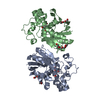
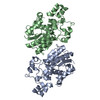
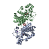
- PDB-3fdz: Crystal structure of phosphoglyceromutase from burkholderia pseud... -
+ Open data

- Basic information
| Entry | Database: PDB / ID: 3fdz | ||||||
|---|---|---|---|---|---|---|---|
| Title | Crystal structure of phosphoglyceromutase from burkholderia pseudomallei 1710b with bound 2,3-diphosphoglyceric acid and 3-phosphoglyceric acid | ||||||
 Components Components | (2,3-bisphosphoglycerate-dependent phosphoglycerate ...) x 2 | ||||||
 Keywords Keywords | ISOMERASE / SSGCID / Phosphoglyceromutase / Burkholderia pseudomallei / Glycolysis / Structural Genomics / Seattle Structural Genomics Center for Infectious Disease | ||||||
| Function / homology |  Function and homology information Function and homology informationphosphoglycerate mutase (2,3-diphosphoglycerate-dependent) / phosphoglycerate mutase activity / gluconeogenesis / glycolytic process Similarity search - Function | ||||||
| Biological species |  Burkholderia pseudomallei 1710b (bacteria) Burkholderia pseudomallei 1710b (bacteria) | ||||||
| Method |  X-RAY DIFFRACTION / MOLECULAR REPLACEMENT / Resolution: 2.25 Å X-RAY DIFFRACTION / MOLECULAR REPLACEMENT / Resolution: 2.25 Å | ||||||
 Authors Authors | Seattle Structural Genomics Center for Infectious Disease (SSGCID) | ||||||
 Citation Citation | Journal: Acta Crystallogr.,Sect.F / Year: 2011 Title: An ensemble of structures of Burkholderia pseudomallei 2,3-bisphosphoglycerate-dependent phosphoglycerate mutase. Authors: Davies, D.R. / Staker, B.L. / Abendroth, J.A. / Edwards, T.E. / Hartley, R. / Leonard, J. / Kim, H. / Rychel, A.L. / Hewitt, S.N. / Myler, P.J. / Stewart, L.J. | ||||||
| History |
|
- Structure visualization
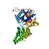
| Structure viewer | Molecule: MolmilJmol/JSmol |
|---|
- Downloads & links
-Download
| PDBx/mmCIF format | 3fdz.cif.gz | 110.3 KB | Display | PDBx/mmCIF format |
|---|---|---|---|---|
| PDB format | pdb3fdz.ent.gz | 84.2 KB | Display | PDB format |
| PDBx/mmJSON format | 3fdz.json.gz | Tree view | PDBx/mmJSON format | |
| Others |  Other downloads Other downloads |
-Validation report
| Arichive directory | https://data.pdbj.org/pub/pdb/validation_reports/fd/3fdzftp://data.pdbj.org/pub/pdb/validation_reports/fd/3fdz | HTTPS FTP |
|---|
-Related structure data
| Related structure data |  3eznC  3gp3C  3gp5C  3gw8C  3lntC C: citing same article ( |
|---|---|
| Similar structure data | |
| Other databases |


-Links
 PDBj
PDBj


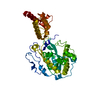
- Assembly
| Deposited unit | 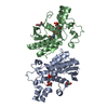
| ||||||||
|---|---|---|---|---|---|---|---|---|---|
| 1 |
| ||||||||
| 2 | 
| ||||||||
| 3 | 
| ||||||||
| Unit cell |
| ||||||||
| Details | The asymmetric unit contains two biological units |
-Components
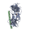
-2,3-bisphosphoglycerate-dependent phosphoglycerate ... , 2 types, 2 molecules AB
| #1: Protein | Mass: 28958.844 Da / Num. of mol.: 1 Source method: isolated from a genetically manipulated source Source: (gene. exp.) Burkholderia pseudomallei 1710b (bacteria)Strain: 1719B / Gene: BURPS1710b_0662, gpmA / Plasmid: AVA0421 / Production host: |
|---|---|
| #2: Protein | Mass: 29037.816 Da / Num. of mol.: 1 Source method: isolated from a genetically manipulated source Source: (gene. exp.) Burkholderia pseudomallei 1710b (bacteria)Strain: 1719B / Gene: BURPS1710b_0662, gpmA / Plasmid: AVA0421 / Production host: |
-Non-polymers , 4 types, 227 molecules 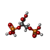


| #3: Chemical | ChemComp-DG2 / ( Mass: 266.037 Da / Num. of mol.: 1 / Source method: obtained synthetically / Formula: C3H8O10P2 Mass: 266.037 Da / Num. of mol.: 1 / Source method: obtained synthetically / Formula: C3H8O10P2 | ||||
|---|---|---|---|---|---|
| #4: Chemical |  Mass: 106.120 Da / Num. of mol.: 2 / Source method: obtained synthetically / Formula: C4H10O3 Mass: 106.120 Da / Num. of mol.: 2 / Source method: obtained synthetically / Formula: C4H10O3#5: Chemical | ChemComp-3PG / |  Mass: 186.057 Da / Num. of mol.: 1 / Source method: obtained synthetically / Formula: C3H7O7P Mass: 186.057 Da / Num. of mol.: 1 / Source method: obtained synthetically / Formula: C3H7O7P#6: Water | ChemComp-HOH / | Mass: 18.015 Da / Num. of mol.: 223 / Source method: isolated from a natural source / Formula: H2O |
-Experimental details
-Experiment
| Experiment | Method: X-RAY DIFFRACTION / Number of used crystals: 1 |
|---|
- Sample preparation
| Crystal | Density Matthews: 2.1 Å3/Da / Density % sol: 41.33 % |
|---|---|
| Crystal grow | Temperature: 289 K / pH: 6 Details: 30% PEG 600, 5% PEG 1000, 10% GLYCEROL, 100 MM MES PH 6.0, CRYSTALS SOAKED OVERNIGHT WITH 20 MM 3-PHOSPHOGLYCERIC ACID, VAPOR DIFFUSION, VAPOR DIFFUSION, SITTING DROP, temperature 289K |
-Data collection
| Diffraction | Mean temperature: 100 K |
|---|---|
| Diffraction source | Source: ROTATING ANODE / Type: RIGAKU / Wavelength: 1.54 |
| Detector | Type: SATURN / Detector: CCD / Date: Sep 30, 2008 |
| Radiation | Protocol: SINGLE WAVELENGTH / Scattering type: x-ray |
| Radiation wavelength | Wavelength: 1.54 Å / Relative weight: 1 |
| Reflection | Highest resolution: 2.25 Å / Num. obs: 20934 / % possible obs: 93.4 % / Observed criterion σ(I): -3 / Biso Wilson estimate: 29.18 Å2 / Rmerge(I) obs: 0.071 / Net I/σ(I): 11.43 |
| Reflection shell | Resolution: 2.25→2.31 Å / Rmerge(I) obs: 0.345 / Mean I/σ(I) obs: 2.6 / % possible all: 90.5 |
- Processing
| Software |
| ||||||||||||||||||||||||||||||||||||||||||||||||||||||||||||||||||||||||||||||||||||||||||||||||||||||||||||||||||||||||||||||||||||||||||||||||||||||||||||||||||||||||||
|---|---|---|---|---|---|---|---|---|---|---|---|---|---|---|---|---|---|---|---|---|---|---|---|---|---|---|---|---|---|---|---|---|---|---|---|---|---|---|---|---|---|---|---|---|---|---|---|---|---|---|---|---|---|---|---|---|---|---|---|---|---|---|---|---|---|---|---|---|---|---|---|---|---|---|---|---|---|---|---|---|---|---|---|---|---|---|---|---|---|---|---|---|---|---|---|---|---|---|---|---|---|---|---|---|---|---|---|---|---|---|---|---|---|---|---|---|---|---|---|---|---|---|---|---|---|---|---|---|---|---|---|---|---|---|---|---|---|---|---|---|---|---|---|---|---|---|---|---|---|---|---|---|---|---|---|---|---|---|---|---|---|---|---|---|---|---|---|---|---|---|---|
| Refinement | Method to determine structure: MOLECULAR REPLACEMENT / Resolution: 2.25→19.75 Å / Cor.coef. Fo:Fc: 0.944 / Cor.coef. Fo:Fc free: 0.891 / SU B: 7.03 / SU ML: 0.174 / Cross valid method: THROUGHOUT / σ(F): 0 / ESU R: 0.418 / ESU R Free: 0.266 / Stereochemistry target values: MAXIMUM LIKELIHOOD / Details: HYDROGENS HAVE BEEN ADDED IN THE
| ||||||||||||||||||||||||||||||||||||||||||||||||||||||||||||||||||||||||||||||||||||||||||||||||||||||||||||||||||||||||||||||||||||||||||||||||||||||||||||||||||||||||||
| Solvent computation | Ion probe radii: 0.8 Å / Shrinkage radii: 0.8 Å / VDW probe radii: 1.2 Å / Solvent model: MASK | ||||||||||||||||||||||||||||||||||||||||||||||||||||||||||||||||||||||||||||||||||||||||||||||||||||||||||||||||||||||||||||||||||||||||||||||||||||||||||||||||||||||||||
| Displacement parameters | Biso mean: 23.7 Å2
| ||||||||||||||||||||||||||||||||||||||||||||||||||||||||||||||||||||||||||||||||||||||||||||||||||||||||||||||||||||||||||||||||||||||||||||||||||||||||||||||||||||||||||
| Refinement step | Cycle: LAST / Resolution: 2.25→19.75 Å
| ||||||||||||||||||||||||||||||||||||||||||||||||||||||||||||||||||||||||||||||||||||||||||||||||||||||||||||||||||||||||||||||||||||||||||||||||||||||||||||||||||||||||||
| Refine LS restraints |
| ||||||||||||||||||||||||||||||||||||||||||||||||||||||||||||||||||||||||||||||||||||||||||||||||||||||||||||||||||||||||||||||||||||||||||||||||||||||||||||||||||||||||||
| LS refinement shell | Resolution: 2.25→2.31 Å / Total num. of bins used: 20
|
