 Movie
Movie Controller

[English] 日本語
 Yorodumi
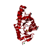
- PDB-5kc8: Crystal structure of the amino-terminal domain (ATD) of iGluR Del... -
+ Open data

- Basic information
| Entry | Database: PDB / ID: 5kc8 | ||||||
|---|---|---|---|---|---|---|---|
| Title | Crystal structure of the amino-terminal domain (ATD) of iGluR Delta-2 (GluD2) | ||||||
 Components Components | Glutamate receptor ionotropic, delta-2 | ||||||
 Keywords Keywords | SIGNALING PROTEIN / ionotropic glutamate receptor (iGluR) / neurotransmission | ||||||
| Function / homology |  Function and homology information Function and homology informationtrans-synaptic signaling by trans-synaptic complex, modulating synaptic transmission / trans-synaptic protein complex / cerebellar granule cell differentiation / positive regulation of long-term synaptic depression / excitatory synapse assembly / synaptic signaling via neuropeptide / regulation of postsynaptic density assembly / positive regulation of synapse assembly / glutamate receptor activity / heterophilic cell-cell adhesion ...trans-synaptic signaling by trans-synaptic complex, modulating synaptic transmission / trans-synaptic protein complex / cerebellar granule cell differentiation / positive regulation of long-term synaptic depression / excitatory synapse assembly / synaptic signaling via neuropeptide / regulation of postsynaptic density assembly / positive regulation of synapse assembly / glutamate receptor activity / heterophilic cell-cell adhesion / glutamate receptor signaling pathway / AMPA glutamate receptor activity / parallel fiber to Purkinje cell synapse / AMPA glutamate receptor complex / ionotropic glutamate receptor complex / regulation of presynapse assembly / regulation of postsynaptic membrane neurotransmitter receptor levels / prepulse inhibition / regulation of neuron apoptotic process / excitatory postsynaptic potential / PDZ domain binding / synaptic transmission, glutamatergic / transmitter-gated monoatomic ion channel activity involved in regulation of postsynaptic membrane potential / postsynaptic density membrane / modulation of chemical synaptic transmission / intracellular protein localization / scaffold protein binding / dendritic spine / synapse / glutamatergic synapse / metal ion binding / identical protein binding / plasma membrane Similarity search - Function | ||||||
| Biological species |  Homo sapiens (human) Homo sapiens (human) | ||||||
| Method |  X-RAY DIFFRACTION / SYNCHROTRON / MOLECULAR REPLACEMENT / Resolution: 1.751 Å X-RAY DIFFRACTION / SYNCHROTRON / MOLECULAR REPLACEMENT / Resolution: 1.751 Å | ||||||
 Authors Authors | Elegheert, J. / Clay, J.E. / Siebold, C. / Aricescu, A.R. | ||||||
 Citation Citation | Journal: Science / Year: 2016 Title: Structural basis for integration of GluD receptors within synaptic organizer complexes. Authors: Elegheert, J. / Kakegawa, W. / Clay, J.E. / Shanks, N.F. / Behiels, E. / Matsuda, K. / Kohda, K. / Miura, E. / Rossmann, M. / Mitakidis, N. / Motohashi, J. / Chang, V.T. / Siebold, C. / ...Authors: Elegheert, J. / Kakegawa, W. / Clay, J.E. / Shanks, N.F. / Behiels, E. / Matsuda, K. / Kohda, K. / Miura, E. / Rossmann, M. / Mitakidis, N. / Motohashi, J. / Chang, V.T. / Siebold, C. / Greger, I.H. / Nakagawa, T. / Yuzaki, M. / Aricescu, A.R. | ||||||
| History |
|
- Structure visualization
| Structure viewer | Molecule: MolmilJmol/JSmol |
|---|
- Downloads & links
-Download
| PDBx/mmCIF format | 5kc8.cif.gz | 100.7 KB | Display | PDBx/mmCIF format |
|---|---|---|---|---|
| PDB format | pdb5kc8.ent.gz | 74.1 KB | Display | PDB format |
| PDBx/mmJSON format | 5kc8.json.gz | Tree view | PDBx/mmJSON format | |
| Others |  Other downloads Other downloads |
-Validation report
| Arichive directory | https://data.pdbj.org/pub/pdb/validation_reports/kc/5kc8ftp://data.pdbj.org/pub/pdb/validation_reports/kc/5kc8 | HTTPS FTP |
|---|
-Related structure data
| Related structure data | 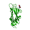 5kc5C  5kc6C 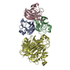 5kc7C 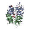 5kc9C 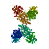 5kcaC  2wjwS S: Starting model for refinement C: citing same article ( |
|---|---|
| Similar structure data |


-Links
 PDBj
PDBj

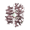
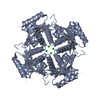
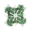
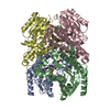
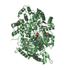
- Assembly
| Deposited unit | 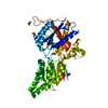
| ||||||||
|---|---|---|---|---|---|---|---|---|---|
| 1 | 
| ||||||||
| Unit cell |
| ||||||||
| Symmetry | Point symmetry: (Schoenflies symbol: C2 (2 fold cyclic)) |
-Components
| #1: Protein | Mass: 48470.797 Da / Num. of mol.: 1 Source method: isolated from a genetically manipulated source Source: (gene. exp.) Homo sapiens (human) / Gene: GRID2, GLURD2 / Plasmid: pHLsec / Cell line (production host): HEK293S / Production host: Homo sapiens (human) / References: UniProt: O43424 | ||||||
|---|---|---|---|---|---|---|---|
| #2: Chemical |   Mass: 40.078 Da / Num. of mol.: 2 / Source method: obtained synthetically / Formula: Ca Mass: 40.078 Da / Num. of mol.: 2 / Source method: obtained synthetically / Formula: Ca#3: Chemical |   Mass: 62.068 Da / Num. of mol.: 3 / Source method: obtained synthetically / Formula: C2H6O2 Mass: 62.068 Da / Num. of mol.: 3 / Source method: obtained synthetically / Formula: C2H6O2#4: Water | ChemComp-HOH / |  Mass: 18.015 Da / Num. of mol.: 237 / Source method: isolated from a natural source / Formula: H2O Mass: 18.015 Da / Num. of mol.: 237 / Source method: isolated from a natural source / Formula: H2OHas protein modification | Y | |
-Experimental details
-Experiment
| Experiment | Method: X-RAY DIFFRACTION / Number of used crystals: 1 |
|---|
- Sample preparation
| Crystal | Density Matthews: 2.27 Å3/Da / Density % sol: 45.84 % |
|---|---|
| Crystal grow | Temperature: 293 K / Method: vapor diffusion, sitting drop Details: 20% (w/v) polyethylene glycol 1000, 0.2 M calcium Acetate, 0.1 M imidazole pH 8.0 and 0.4 M NDSB-221 (non-detergent sulfobetaine 221) |
-Data collection
| Diffraction | Mean temperature: 100 K |
|---|---|
| Diffraction source | Source: SYNCHROTRON / Site: Diamond  / Beamline: I04 / Wavelength: 1 Å / Beamline: I04 / Wavelength: 1 Å |
| Detector | Type: ADSC QUANTUM 315 / Detector: CCD / Date: Jun 27, 2010 |
| Radiation | Protocol: SINGLE WAVELENGTH / Monochromatic (M) / Laue (L): M / Scattering type: x-ray |
| Radiation wavelength | Wavelength: 1 Å / Relative weight: 1 |
| Reflection | Resolution: 1.75→57.03 Å / Num. obs: 44958 / % possible obs: 99.6 % / Redundancy: 15.3 % / Biso Wilson estimate: 25.6 Å2 / CC1/2: 1 / Rmerge(I) obs: 0.078 / Net I/σ(I): 24.2 |
| Reflection shell | Resolution: 1.75→1.8 Å / Redundancy: 9.5 % / Rmerge(I) obs: 1.314 / Mean I/σ(I) obs: 2.2 / % possible all: 98.3 |
- Processing
| Software |
| |||||||||||||||||||||||||||||||||||||||||||||||||||||||||||||||||||||||||||||||||||||||||||||||||||||||||||||||||||||||
|---|---|---|---|---|---|---|---|---|---|---|---|---|---|---|---|---|---|---|---|---|---|---|---|---|---|---|---|---|---|---|---|---|---|---|---|---|---|---|---|---|---|---|---|---|---|---|---|---|---|---|---|---|---|---|---|---|---|---|---|---|---|---|---|---|---|---|---|---|---|---|---|---|---|---|---|---|---|---|---|---|---|---|---|---|---|---|---|---|---|---|---|---|---|---|---|---|---|---|---|---|---|---|---|---|---|---|---|---|---|---|---|---|---|---|---|---|---|---|---|---|
| Refinement | Method to determine structure: MOLECULAR REPLACEMENT Starting model: 2WJW Resolution: 1.751→41.871 Å / SU ML: 0.21 / Cross valid method: FREE R-VALUE / σ(F): 1.34 / Phase error: 25.44 / Stereochemistry target values: ML
| |||||||||||||||||||||||||||||||||||||||||||||||||||||||||||||||||||||||||||||||||||||||||||||||||||||||||||||||||||||||
| Solvent computation | Shrinkage radii: 1 Å / VDW probe radii: 1.2 Å / Solvent model: FLAT BULK SOLVENT MODEL | |||||||||||||||||||||||||||||||||||||||||||||||||||||||||||||||||||||||||||||||||||||||||||||||||||||||||||||||||||||||
| Displacement parameters | Biso mean: 41.8 Å2 | |||||||||||||||||||||||||||||||||||||||||||||||||||||||||||||||||||||||||||||||||||||||||||||||||||||||||||||||||||||||
| Refinement step | Cycle: LAST / Resolution: 1.751→41.871 Å
| |||||||||||||||||||||||||||||||||||||||||||||||||||||||||||||||||||||||||||||||||||||||||||||||||||||||||||||||||||||||
| Refine LS restraints |
| |||||||||||||||||||||||||||||||||||||||||||||||||||||||||||||||||||||||||||||||||||||||||||||||||||||||||||||||||||||||
| LS refinement shell |
|
