 Movie
Movie Controller

[English] 日本語
 Yorodumi
- PDB-6ds8: Crystal structure of MhuD R26S mutant with two Manganese protopor... -
+ Open data

- Basic information
| Entry | Database: PDB / ID: 6ds8 | |||||||||
|---|---|---|---|---|---|---|---|---|---|---|
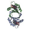
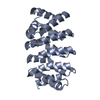
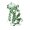
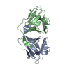
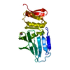
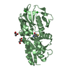
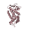
| Title | Crystal structure of MhuD R26S mutant with two Manganese protoporphyrin IX bound per active site | |||||||||
 Components Components | Heme-degrading monooxygenase HmoB | |||||||||
 Keywords Keywords | OXIDOREDUCTASE / Heme Oxygenase (decyclizing) Activity / Monooxygenase Activity / Oxidoreductase Activity / Heme Binding / Metal Ion Binding / Heme Catabolic Process / Oxidation Reduction Process / Cell Wall / Plasma Membrane | |||||||||
| Function / homology |  Function and homology information Function and homology informationheme oxygenase (mycobilin-producing) / heme oxygenase (decyclizing) activity / heme catabolic process / peptidoglycan-based cell wall / heme binding / metal ion binding / plasma membrane Similarity search - Function | |||||||||
| Biological species |   Mycobacterium tuberculosis (bacteria) Mycobacterium tuberculosis (bacteria) | |||||||||
| Method |  X-RAY DIFFRACTION / SYNCHROTRON / MOLECULAR REPLACEMENT / Resolution: 2.4 Å X-RAY DIFFRACTION / SYNCHROTRON / MOLECULAR REPLACEMENT / Resolution: 2.4 Å | |||||||||
 Authors Authors | Chao, A. / Goulding, C.W. | |||||||||
| Funding support |  United States, 2items United States, 2items
| |||||||||
 Citation Citation | Journal: To Be Published Title: Crystal structure of MhuD R26S mutant with two Manganese protoporphyrin IX bound per active site Authors: Chao, A. / Goulding, C.W. | |||||||||
| History |
|
- Structure visualization
| Structure viewer | Molecule: MolmilJmol/JSmol |
|---|
- Downloads & links
-Download
| PDBx/mmCIF format | 6ds8.cif.gz | 57.7 KB | Display | PDBx/mmCIF format |
|---|---|---|---|---|
| PDB format | pdb6ds8.ent.gz | 40.1 KB | Display | PDB format |
| PDBx/mmJSON format | 6ds8.json.gz | Tree view | PDBx/mmJSON format | |
| Others |  Other downloads Other downloads |
-Validation report
| Arichive directory | https://data.pdbj.org/pub/pdb/validation_reports/ds/6ds8ftp://data.pdbj.org/pub/pdb/validation_reports/ds/6ds8 | HTTPS FTP |
|---|
-Related structure data
| Related structure data |  3hx9S S: Starting model for refinement |
|---|---|
| Similar structure data |


-Links
 PDBj
PDBj- Assembly
| Deposited unit | 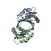
| |||||||||||||||||||||
|---|---|---|---|---|---|---|---|---|---|---|---|---|---|---|---|---|---|---|---|---|---|---|
| 1 |
| |||||||||||||||||||||
| Unit cell |
| |||||||||||||||||||||
| Noncrystallographic symmetry (NCS) | NCS domain:
NCS domain segments: Component-ID: 1 / Ens-ID: 1 / Beg auth comp-ID: PRO / Beg label comp-ID: PRO / End auth comp-ID: GLY / End label comp-ID: GLY / Auth seq-ID: 2 - 100 / Label seq-ID: 1 - 99
|
-Components
| #1: Protein | Mass: 10537.784 Da / Num. of mol.: 2 / Mutation: R26S Source method: isolated from a genetically manipulated source Source: (gene. exp.) Mycobacterium tuberculosis (strain ATCC 25618 / H37Rv) (bacteria)Strain: ATCC 25618 / H37Rv / Gene: mhuD, Rv3592 / Production host: References: UniProt: P9WKH3, heme oxygenase (biliverdin-producing) #2: Chemical | ChemComp-MNH /   Mass: 615.580 Da / Num. of mol.: 4 / Source method: obtained synthetically / Formula: C34H32MnN4O4 / Feature type: SUBJECT OF INVESTIGATION Mass: 615.580 Da / Num. of mol.: 4 / Source method: obtained synthetically / Formula: C34H32MnN4O4 / Feature type: SUBJECT OF INVESTIGATION#3: Chemical |   Mass: 35.453 Da / Num. of mol.: 2 / Source method: obtained synthetically / Formula: Cl Mass: 35.453 Da / Num. of mol.: 2 / Source method: obtained synthetically / Formula: Cl#4: Water | ChemComp-HOH / |  Mass: 18.015 Da / Num. of mol.: 24 / Source method: isolated from a natural source / Formula: H2O Mass: 18.015 Da / Num. of mol.: 24 / Source method: isolated from a natural source / Formula: H2O |
|---|
-Experimental details
-Experiment
| Experiment | Method: X-RAY DIFFRACTION / Number of used crystals: 1 |
|---|
- Sample preparation
| Crystal | Density Matthews: 2.37 Å3/Da / Density % sol: 48.07 % |
|---|---|
| Crystal grow | Temperature: 298 K / Method: vapor diffusion, hanging drop / pH: 7.5 Details: 0.2 M ammonium sulfate, 0.1 M HEPES pH 7.5, 25% (w/v) Polyethylene glycol 3350 |
-Data collection
| Diffraction | Mean temperature: 100 K |
|---|---|
| Diffraction source | Source: SYNCHROTRON / Site: SSRL / Beamline: BL14-1 / Wavelength: 1 Å |
| Detector | Type: MARMOSAIC 325 mm CCD / Detector: CCD / Date: Jul 7, 2016 |
| Radiation | Protocol: SINGLE WAVELENGTH / Monochromatic (M) / Laue (L): M / Scattering type: x-ray |
| Radiation wavelength | Wavelength: 1 Å / Relative weight: 1 |
| Reflection | Resolution: 2.4→50.502 Å / Num. obs: 7868 / % possible obs: 95.06 % / Redundancy: 2 % / Biso Wilson estimate: 30.69 Å2 / CC1/2: 0.999 / Rmerge(I) obs: 0.01736 / Rpim(I) all: 0.01736 / Rrim(I) all: 0.02455 / Net I/σ(I): 26.25 |
| Reflection shell | Resolution: 2.4→2.486 Å / Rmerge(I) obs: 0.08277 / Mean I/σ(I) obs: 7.58 / Num. unique obs: 760 / CC1/2: 0.98 / Rpim(I) all: 0.08277 / Rrim(I) all: 0.1171 / % possible all: 93.6 |
- Processing
| Software |
| |||||||||||||||||||||||||||||||||||||||||||||||||
|---|---|---|---|---|---|---|---|---|---|---|---|---|---|---|---|---|---|---|---|---|---|---|---|---|---|---|---|---|---|---|---|---|---|---|---|---|---|---|---|---|---|---|---|---|---|---|---|---|---|---|
| Refinement | Method to determine structure: MOLECULAR REPLACEMENT Starting model: 3HX9 Resolution: 2.4→50.502 Å / SU ML: 0.28 / Cross valid method: THROUGHOUT / σ(F): 1.36 / Phase error: 23.25
| |||||||||||||||||||||||||||||||||||||||||||||||||
| Solvent computation | Shrinkage radii: 0.9 Å / VDW probe radii: 1.11 Å | |||||||||||||||||||||||||||||||||||||||||||||||||
| Displacement parameters | Biso max: 80.26 Å2 / Biso mean: 30.6722 Å2 / Biso min: 10.77 Å2 | |||||||||||||||||||||||||||||||||||||||||||||||||
| Refinement step | Cycle: final / Resolution: 2.4→50.502 Å
| |||||||||||||||||||||||||||||||||||||||||||||||||
| Refine LS restraints NCS |
| |||||||||||||||||||||||||||||||||||||||||||||||||
| LS refinement shell | Refine-ID: X-RAY DIFFRACTION / Rfactor Rfree error: 0 / Total num. of bins used: 6
|
