 Movie
Movie Controller

[English] 日本語
 Yorodumi
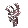
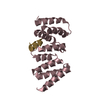
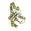
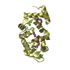
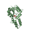
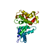
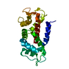
- PDB-3hx9: Structure of heme-degrader, MhuD (Rv3592), from Mycobacterium tub... -
+ Open data

- Basic information
| Entry | Database: PDB / ID: 3hx9 | ||||||
|---|---|---|---|---|---|---|---|
| Title | Structure of heme-degrader, MhuD (Rv3592), from Mycobacterium tuberculosis with two hemes bound in its active site | ||||||
 Components Components | Protein Rv3592 | ||||||
 Keywords Keywords | OXIDOREDUCTASE / di-heme / beta barrel / dimer | ||||||
| Function / homology |  Function and homology information Function and homology informationheme oxygenase (mycobilin-producing) / cell wall / heme oxygenase (decyclizing) activity / heme catabolic process / peptidoglycan-based cell wall / heme binding / metal ion binding / plasma membrane Similarity search - Function | ||||||
| Biological species |   Mycobacterium tuberculosis (bacteria) Mycobacterium tuberculosis (bacteria) | ||||||
| Method |  X-RAY DIFFRACTION / SYNCHROTRON / MOLECULAR REPLACEMENT / Resolution: 1.75 Å X-RAY DIFFRACTION / SYNCHROTRON / MOLECULAR REPLACEMENT / Resolution: 1.75 Å | ||||||
 Authors Authors | Chim, N. / Nguyen, T.Q. / Iniguez, A. / Goulding, C.W. | ||||||
 Citation Citation | Journal: J.Mol.Biol. / Year: 2009 Title: Unusual Diheme Conformation of the Heme-Degrading Protein from Mycobacterium tuberculosis Authors: Chim, N. / Iniguez, A. / Nguyen, T.Q. / Goulding, C.W. | ||||||
| History |
|
- Structure visualization
| Structure viewer | Molecule: MolmilJmol/JSmol |
|---|
- Downloads & links
-Download
| PDBx/mmCIF format | 3hx9.cif.gz | 58.8 KB | Display | PDBx/mmCIF format |
|---|---|---|---|---|
| PDB format | pdb3hx9.ent.gz | 43.2 KB | Display | PDB format |
| PDBx/mmJSON format | 3hx9.json.gz | Tree view | PDBx/mmJSON format | |
| Others |  Other downloads Other downloads |
-Validation report
| Arichive directory | https://data.pdbj.org/pub/pdb/validation_reports/hx/3hx9ftp://data.pdbj.org/pub/pdb/validation_reports/hx/3hx9 | HTTPS FTP |
|---|
-Related structure data
| Similar structure data |
|---|


-Links
 PDBj
PDBj- Assembly
| Deposited unit | 
| ||||||||
|---|---|---|---|---|---|---|---|---|---|
| 1 |
| ||||||||
| Unit cell |
|
-Components
| #1: Protein | Mass: 13249.933 Da / Num. of mol.: 2 Source method: isolated from a genetically manipulated source Source: (gene. exp.) Mycobacterium tuberculosis (bacteria) / Strain: H37Rv / Gene: MT3698, Rv3592, TB11.2 / Plasmid: pET22b / Production host: #2: Chemical | ChemComp-HEM /   Mass: 616.487 Da / Num. of mol.: 4 / Source method: obtained synthetically / Formula: C34H32FeN4O4 Mass: 616.487 Da / Num. of mol.: 4 / Source method: obtained synthetically / Formula: C34H32FeN4O4#3: Chemical |   Mass: 35.453 Da / Num. of mol.: 2 / Source method: obtained synthetically / Formula: Cl Mass: 35.453 Da / Num. of mol.: 2 / Source method: obtained synthetically / Formula: Cl#4: Water | ChemComp-HOH / |  Mass: 18.015 Da / Num. of mol.: 123 / Source method: isolated from a natural source / Formula: H2O Mass: 18.015 Da / Num. of mol.: 123 / Source method: isolated from a natural source / Formula: H2O |
|---|
-Experimental details
-Experiment
| Experiment | Method: X-RAY DIFFRACTION / Number of used crystals: 1 |
|---|
- Sample preparation
| Crystal | Density Matthews: 1.91 Å3/Da / Density % sol: 35.45 % |
|---|---|
| Crystal grow | Temperature: 298 K / Method: vapor diffusion, hanging drop / pH: 5 Details: 0.1 M Bis-Tris pH 5.0, 0.2 M NaCl, 20% PEG-3350, 10 mM triethylamine HCl , VAPOR DIFFUSION, HANGING DROP, temperature 298K |
-Data collection
| Diffraction | Mean temperature: 70 K | |||||||||
|---|---|---|---|---|---|---|---|---|---|---|
| Diffraction source | Source: SYNCHROTRON / Site: SSRL  / Beamline: BL7-1 / Wavelength: 0.97, 1.76 / Beamline: BL7-1 / Wavelength: 0.97, 1.76 | |||||||||
| Detector | Type: ADSC QUANTUM 315r / Detector: CCD / Date: Dec 7, 2008 Details: Vertical focusing mirror; single crystal (Si111) bent monochromator (horizontal focusing). | |||||||||
| Radiation | Monochromator: side scattering I-beam bent single crystal; asymmetric cut 4.9650 deg. Protocol: SINGLE WAVELENGTH / Monochromatic (M) / Laue (L): M / Scattering type: x-ray | |||||||||
| Radiation wavelength |
| |||||||||
| Reflection | Resolution: 1.75→50 Å / Num. obs: 19968 / % possible obs: 98.9 % / Rmerge(I) obs: 0.066 / Net I/σ(I): 28.1 | |||||||||
| Reflection shell | Resolution: 1.75→1.81 Å / Rmerge(I) obs: 0.165 / Mean I/σ(I) obs: 10.7 / Num. unique all: 1974 / % possible all: 98.6 |
- Processing
| Software |
| |||||||||||||||||||||||||||||||||||||||||||||||||||||||||||||||||
|---|---|---|---|---|---|---|---|---|---|---|---|---|---|---|---|---|---|---|---|---|---|---|---|---|---|---|---|---|---|---|---|---|---|---|---|---|---|---|---|---|---|---|---|---|---|---|---|---|---|---|---|---|---|---|---|---|---|---|---|---|---|---|---|---|---|---|
| Refinement | Method to determine structure: MOLECULAR REPLACEMENT / Resolution: 1.75→19.35 Å / Cor.coef. Fo:Fc: 0.953 / Cor.coef. Fo:Fc free: 0.933 / SU B: 2.34 / SU ML: 0.078 / Cross valid method: THROUGHOUT / ESU R: 0.13 / ESU R Free: 0.127 / Stereochemistry target values: MAXIMUM LIKELIHOOD / Details: HYDROGENS HAVE BEEN ADDED IN THE RIDING POSITIONS
| |||||||||||||||||||||||||||||||||||||||||||||||||||||||||||||||||
| Solvent computation | Ion probe radii: 0.8 Å / Shrinkage radii: 0.8 Å / VDW probe radii: 1.4 Å / Solvent model: MASK | |||||||||||||||||||||||||||||||||||||||||||||||||||||||||||||||||
| Displacement parameters | Biso mean: 22.804 Å2
| |||||||||||||||||||||||||||||||||||||||||||||||||||||||||||||||||
| Refinement step | Cycle: LAST / Resolution: 1.75→19.35 Å
| |||||||||||||||||||||||||||||||||||||||||||||||||||||||||||||||||
| Refine LS restraints |
| |||||||||||||||||||||||||||||||||||||||||||||||||||||||||||||||||
| LS refinement shell | Resolution: 1.749→1.794 Å / Total num. of bins used: 20
|
