 Movie
Movie Controller

[English] 日本語
 Yorodumi
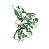
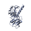
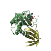
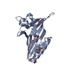
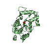
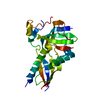
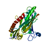
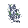
- PDB-3p9y: Crystal structure of the Drosophila melanogaster Ssu72-pCTD complex -
+ Open data

- Basic information
| Entry | Database: PDB / ID: 3p9y | ||||||
|---|---|---|---|---|---|---|---|
| Title | Crystal structure of the Drosophila melanogaster Ssu72-pCTD complex | ||||||
 Components Components |
| ||||||
 Keywords Keywords | HYDROLASE / Phosphatase / cis proline / LMW PTP-like fold / RNA polymerase II CTD | ||||||
| Function / homology |  Function and homology information Function and homology informationRNA polymerase II transcribes snRNA genes / RNA polymerase II CTD heptapeptide repeat phosphatase activity / mRNA cleavage and polyadenylation specificity factor complex / mRNA 3'-end processing / protein-serine/threonine phosphatase / termination of RNA polymerase II transcription / phosphatase activity / nucleus Similarity search - Function | ||||||
| Biological species |  synthetic construct (others) | ||||||
| Method |  X-RAY DIFFRACTION / SYNCHROTRON / MOLECULAR REPLACEMENT / Resolution: 2.1 Å X-RAY DIFFRACTION / SYNCHROTRON / MOLECULAR REPLACEMENT / Resolution: 2.1 Å | ||||||
 Authors Authors | Werner-Allen, J.W. / Zhou, P. | ||||||
 Citation Citation | Journal: J.Biol.Chem. / Year: 2011 Title: cis-Proline-mediated Ser(P)5 Dephosphorylation by the RNA Polymerase II C-terminal Domain Phosphatase Ssu72. Authors: Werner-Allen, J.W. / Lee, C.J. / Liu, P. / Nicely, N.I. / Wang, S. / Greenleaf, A.L. / Zhou, P. | ||||||
| History |
|
- Structure visualization
| Structure viewer | Molecule: MolmilJmol/JSmol |
|---|
- Downloads & links
-Download
| PDBx/mmCIF format | 3p9y.cif.gz | 325.2 KB | Display | PDBx/mmCIF format |
|---|---|---|---|---|
| PDB format | pdb3p9y.ent.gz | 270 KB | Display | PDB format |
| PDBx/mmJSON format | 3p9y.json.gz | Tree view | PDBx/mmJSON format | |
| Others |  Other downloads Other downloads |
-Validation report
| Arichive directory | https://data.pdbj.org/pub/pdb/validation_reports/p9/3p9yftp://data.pdbj.org/pub/pdb/validation_reports/p9/3p9y | HTTPS FTP |
|---|
-Related structure data
| Similar structure data |
|---|


-Links
 PDBj
PDBj


- Assembly
| Deposited unit | 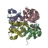
| ||||||||
|---|---|---|---|---|---|---|---|---|---|
| 1 | 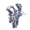
| ||||||||
| 2 | 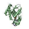
| ||||||||
| 3 | 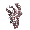
| ||||||||
| 4 | 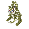
| ||||||||
| Unit cell |
|
-Components
| #1: Protein | Mass: 23057.209 Da / Num. of mol.: 4 / Mutation: C13D,D144N Source method: isolated from a genetically manipulated source Source: (gene. exp.)  #2: Protein/peptide | Mass: 858.787 Da / Num. of mol.: 4 / Source method: obtained synthetically / Details: synthetic phosphopeptide / Source: (synth.) synthetic construct (others) #3: Chemical | ChemComp-IMD /   Mass: 69.085 Da / Num. of mol.: 4 / Source method: obtained synthetically / Formula: C3H5N2 Mass: 69.085 Da / Num. of mol.: 4 / Source method: obtained synthetically / Formula: C3H5N2#4: Chemical | ChemComp-PG4 / |   Mass: 194.226 Da / Num. of mol.: 1 / Source method: obtained synthetically / Formula: C8H18O5 / Comment: precipitant*YM Mass: 194.226 Da / Num. of mol.: 1 / Source method: obtained synthetically / Formula: C8H18O5 / Comment: precipitant*YM#5: Water | ChemComp-HOH / |  Mass: 18.015 Da / Num. of mol.: 658 / Source method: isolated from a natural source / Formula: H2O Mass: 18.015 Da / Num. of mol.: 658 / Source method: isolated from a natural source / Formula: H2OHas protein modification | Y | |
|---|
-Experimental details
-Experiment
| Experiment | Method: X-RAY DIFFRACTION / Number of used crystals: 1 |
|---|
- Sample preparation
| Crystal | Density Matthews: 2.96 Å3/Da / Density % sol: 58.48 % |
|---|---|
| Crystal grow | Temperature: 277 K / Method: vapor diffusion, hanging drop / pH: 6.5 Details: 22% PEG monomethyl ether 550, 100 mM imidazole pH 6.5, 150 mM DL-malic acid, VAPOR DIFFUSION, HANGING DROP, temperature 277K |
-Data collection
| Diffraction | Mean temperature: 100 K |
|---|---|
| Diffraction source | Source: SYNCHROTRON / Site: APS  / Beamline: 22-ID / Wavelength: 1 Å / Beamline: 22-ID / Wavelength: 1 Å |
| Detector | Type: MARMOSAIC 300 mm CCD / Detector: CCD / Date: Nov 27, 2009 |
| Radiation | Monochromator: Si(111) / Protocol: SINGLE WAVELENGTH / Monochromatic (M) / Laue (L): M / Scattering type: x-ray |
| Radiation wavelength | Wavelength: 1 Å / Relative weight: 1 |
| Reflection | Resolution: 2.1→50 Å / Num. all: 63987 / Num. obs: 63898 / % possible obs: 99.9 % / Redundancy: 3.9 % / Rmerge(I) obs: 0.061 |
| Reflection shell | Resolution: 2.1→2.18 Å / Redundancy: 3.9 % / Rmerge(I) obs: 0.326 / Mean I/σ(I) obs: 4.1 / % possible all: 99.8 |
- Processing
| Software |
| ||||||||||||||||||||||||
|---|---|---|---|---|---|---|---|---|---|---|---|---|---|---|---|---|---|---|---|---|---|---|---|---|---|
| Refinement | Method to determine structure: MOLECULAR REPLACEMENT / Resolution: 2.1→36 Å / SU ML: 0.29 / σ(F): 0.11 / Stereochemistry target values: ML
| ||||||||||||||||||||||||
| Solvent computation | Shrinkage radii: 0.9 Å / VDW probe radii: 1.11 Å / Solvent model: FLAT BULK SOLVENT MODEL / Bsol: 41.352 Å2 / ksol: 0.399 e/Å3 | ||||||||||||||||||||||||
| Displacement parameters |
| ||||||||||||||||||||||||
| Refinement step | Cycle: LAST / Resolution: 2.1→36 Å
| ||||||||||||||||||||||||
| Refine LS restraints |
| ||||||||||||||||||||||||
| LS refinement shell | Resolution: 2.1015→2.1766 Å
|
