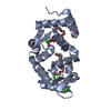
 Movie
Movie Controller


+ Open data

- Basic information
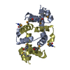
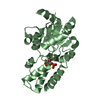
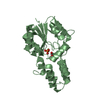
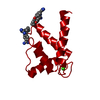
| Entry | Database: PDB / ID: 4guk | ||||||
|---|---|---|---|---|---|---|---|
| Title | New crystal form structure of human NCS1 | ||||||
 Components Components | Neuronal calcium sensor 1 | ||||||
 Keywords Keywords | PROTEIN BINDING / alpha / neuronal calcium sensor | ||||||
| Function / homology |  Function and homology information Function and homology informationcalcium sensitive guanylate cyclase activator activity / regulation of neuron projection development / regulation of signal transduction / voltage-gated calcium channel activity / postsynaptic density / axon / calcium ion binding / dendrite / perinuclear region of cytoplasm / Golgi apparatus ...calcium sensitive guanylate cyclase activator activity / regulation of neuron projection development / regulation of signal transduction / voltage-gated calcium channel activity / postsynaptic density / axon / calcium ion binding / dendrite / perinuclear region of cytoplasm / Golgi apparatus / plasma membrane / cytosol / cytoplasm Similarity search - Function | ||||||
| Biological species |  Homo sapiens (human) Homo sapiens (human) | ||||||
| Method |  X-RAY DIFFRACTION / SYNCHROTRON / MOLECULAR REPLACEMENT / Resolution: 1.75 Å X-RAY DIFFRACTION / SYNCHROTRON / MOLECULAR REPLACEMENT / Resolution: 1.75 Å | ||||||
 Authors Authors | Fan, C. / Lolis, E. | ||||||
 Citation Citation | Journal: To be Published Title: new crystal form structure of human NCS1 Authors: Fan, C. / Lolis, E. | ||||||
| History |
|
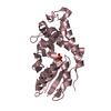
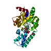
- Structure visualization
| Structure viewer | Molecule: MolmilJmol/JSmol |
|---|
- Downloads & links
-Download
| PDBx/mmCIF format | 4guk.cif.gz | 318.1 KB | Display | PDBx/mmCIF format |
|---|---|---|---|---|
| PDB format | pdb4guk.ent.gz | 255.9 KB | Display | PDB format |
| PDBx/mmJSON format | 4guk.json.gz | Tree view | PDBx/mmJSON format | |
| Others |  Other downloads Other downloads |
-Validation report
| Arichive directory | https://data.pdbj.org/pub/pdb/validation_reports/gu/4gukftp://data.pdbj.org/pub/pdb/validation_reports/gu/4guk | HTTPS FTP |
|---|
-Related structure data
| Similar structure data |
|---|
-Links
 PDBj
PDBj- Assembly
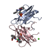
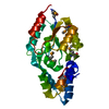
| Deposited unit | 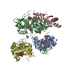
| ||||||||
|---|---|---|---|---|---|---|---|---|---|
| 1 | 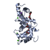
| ||||||||
| 2 | 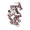
| ||||||||
| 3 | 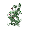
| ||||||||
| 4 | 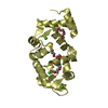
| ||||||||
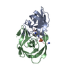
| Unit cell |
|
-Components
-Protein , 1 types, 4 molecules ACBD
| #1: Protein | Mass: 21902.668 Da / Num. of mol.: 4 Source method: isolated from a genetically manipulated source Details: 0.1mMIPTG 16C induction / Source: (gene. exp.) Homo sapiens (human) / Strain: human / Gene: FLUP, FREQ, NCS1 / Plasmid: pET21a(+) / Production host:  |
|---|
-Non-polymers , 8 types, 408 molecules 


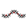
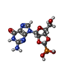


| #2: Chemical | ChemComp-PG4 /  Mass: 194.226 Da / Num. of mol.: 7 / Source method: obtained synthetically / Formula: C8H18O5 / Comment: precipitant*YM Mass: 194.226 Da / Num. of mol.: 7 / Source method: obtained synthetically / Formula: C8H18O5 / Comment: precipitant*YM#3: Chemical | ChemComp-EDO /  Mass: 62.068 Da / Num. of mol.: 5 / Source method: obtained synthetically / Formula: C2H6O2 Mass: 62.068 Da / Num. of mol.: 5 / Source method: obtained synthetically / Formula: C2H6O2#4: Chemical | ChemComp-CA /  Mass: 40.078 Da / Num. of mol.: 12 / Source method: obtained synthetically / Formula: Ca Mass: 40.078 Da / Num. of mol.: 12 / Source method: obtained synthetically / Formula: Ca#5: Chemical | ChemComp-NA /  Mass: 22.990 Da / Num. of mol.: 4 / Source method: obtained synthetically / Formula: Na Mass: 22.990 Da / Num. of mol.: 4 / Source method: obtained synthetically / Formula: Na#6: Chemical | ChemComp-P3G / |  Mass: 250.332 Da / Num. of mol.: 1 / Source method: obtained synthetically / Formula: C12H26O5 Mass: 250.332 Da / Num. of mol.: 1 / Source method: obtained synthetically / Formula: C12H26O5#7: Chemical | ChemComp-P2G / (  Mass: 375.231 Da / Num. of mol.: 4 / Source method: obtained synthetically / Formula: C11H14N5O8P Mass: 375.231 Da / Num. of mol.: 4 / Source method: obtained synthetically / Formula: C11H14N5O8P#8: Chemical | ChemComp-ALA / |  Type: L-peptide linking / Mass: 89.093 Da / Num. of mol.: 1 / Source method: obtained synthetically / Formula: C3H7NO2 Type: L-peptide linking / Mass: 89.093 Da / Num. of mol.: 1 / Source method: obtained synthetically / Formula: C3H7NO2#9: Water | ChemComp-HOH / | Mass: 18.015 Da / Num. of mol.: 374 / Source method: isolated from a natural source / Formula: H2O |
|---|
-Experimental details
-Experiment
| Experiment | Method: X-RAY DIFFRACTION / Number of used crystals: 1 |
|---|
- Sample preparation
| Crystal | Density Matthews: 2.13 Å3/Da / Density % sol: 42.29 % |
|---|---|
| Crystal grow | Temperature: 277 K / Method: vapor diffusion, hanging drop / pH: 6.5 Details: pH6.5, 0.2M NaAC, 30%PEG8000, VAPOR DIFFUSION, HANGING DROP, temperature 277K |
-Data collection
| Diffraction | Mean temperature: 100 K |
|---|---|
| Diffraction source | Source: SYNCHROTRON / Site: NSLS  / Beamline: X25 / Wavelength: 1.1 Å / Beamline: X25 / Wavelength: 1.1 Å |
| Detector | Type: PSI PILATUS 6M / Detector: PIXEL / Date: Apr 28, 2011 |
| Radiation | Monochromator: graphite / Protocol: SINGLE WAVELENGTH / Monochromatic (M) / Laue (L): M / Scattering type: x-ray |
| Radiation wavelength | Wavelength: 1.1 Å / Relative weight: 1 |
| Reflection | Resolution: 1.75→50 Å / Num. all: 75302 / Num. obs: 73721 / % possible obs: 97.9 % / Observed criterion σ(F): 2.7 / Observed criterion σ(I): 2.7 / Redundancy: 7.3 % / Rmerge(I) obs: 0.091 / Rsym value: 0.091 / Net I/σ(I): 11 |
- Processing
| Software |
| |||||||||||||||||||||||||
|---|---|---|---|---|---|---|---|---|---|---|---|---|---|---|---|---|---|---|---|---|---|---|---|---|---|---|
| Refinement | Method to determine structure: MOLECULAR REPLACEMENT Starting model: human NCS1 Resolution: 1.75→50 Å / Isotropic thermal model: isotropic / Cross valid method: THROUGHOUT / σ(F): 0 / σ(I): 0 / Stereochemistry target values: Engh & Huber
| |||||||||||||||||||||||||
| Refinement step | Cycle: LAST / Resolution: 1.75→50 Å
| |||||||||||||||||||||||||
| Refine LS restraints |
|
