 Movie
Movie Controller

[English] 日本語
 Yorodumi
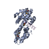
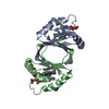
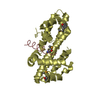
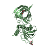
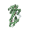
- PDB-3a0c: Crystal Structure of an anti-HIV mannose-binding lectin from Poly... -
+ Open data

- Basic information
| Entry | Database: PDB / ID: 3a0c | ||||||
|---|---|---|---|---|---|---|---|
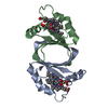
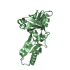
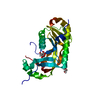
| Title | Crystal Structure of an anti-HIV mannose-binding lectin from Polygonatum cyrtonema Hua | ||||||
 Components Components | Mannose/sialic acid-binding lectin | ||||||
 Keywords Keywords | SUGAR BINDING PROTEIN / beta-prism II / Lectin | ||||||
| Function / homology |  Function and homology information Function and homology information | ||||||
| Biological species |  Polygonatum cyrtonema (plant) Polygonatum cyrtonema (plant) | ||||||
| Method |  X-RAY DIFFRACTION / SYNCHROTRON / MOLECULAR REPLACEMENT / Resolution: 2 Å X-RAY DIFFRACTION / SYNCHROTRON / MOLECULAR REPLACEMENT / Resolution: 2 Å | ||||||
 Authors Authors | Ding, J. / Wang, D.C. | ||||||
 Citation Citation | Journal: J.Struct.Biol. / Year: 2010 Title: Crystal structures of a novel anti-HIV mannose-binding lectin from Polygonatum cyrtonema Hua with unique ligand-binding property and super-structure Authors: Ding, J. / Bao, J. / Zhu, D. / Zhang, Y. / Wang, D.C. | ||||||
| History |
|
- Structure visualization
| Structure viewer | Molecule: MolmilJmol/JSmol |
|---|
- Downloads & links
-Download
| PDBx/mmCIF format | 3a0c.cif.gz | 94.1 KB | Display | PDBx/mmCIF format |
|---|---|---|---|---|
| PDB format | pdb3a0c.ent.gz | 72.9 KB | Display | PDB format |
| PDBx/mmJSON format | 3a0c.json.gz | Tree view | PDBx/mmJSON format | |
| Others |  Other downloads Other downloads |
-Validation report
| Arichive directory | https://data.pdbj.org/pub/pdb/validation_reports/a0/3a0cftp://data.pdbj.org/pub/pdb/validation_reports/a0/3a0c | HTTPS FTP |
|---|
-Related structure data
| Related structure data |  3a0dC 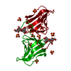 3a0eC 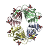 1msaS C: citing same article ( S: Starting model for refinement |
|---|---|
| Similar structure data |

-Links
 PDBj
PDBj
- Assembly
| Deposited unit | 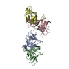
| ||||||||
|---|---|---|---|---|---|---|---|---|---|
| 1 | 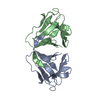
| ||||||||
| 2 | 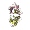
| ||||||||
| Unit cell |
|
-Components
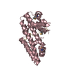
| #1: Protein | Mass: 11962.332 Da / Num. of mol.: 4 / Fragment: UNP residues 29-138 / Source method: isolated from a natural source / Source: (natural) Polygonatum cyrtonema (plant) / Strain: Hua / References: UniProt: Q8L568#2: Water | ChemComp-HOH / |  Mass: 18.015 Da / Num. of mol.: 101 / Source method: isolated from a natural source / Formula: H2O Mass: 18.015 Da / Num. of mol.: 101 / Source method: isolated from a natural source / Formula: H2OHas protein modification | Y | Sequence details | THE CONFLICT BETWEEN THE SEQUENCE OF THE PROTEIN AND DATABASE (UNP Q8L568; Q8L568_9ASP) MAY BE ...THE CONFLICT BETWEEN THE SEQUENCE OF THE PROTEIN AND DATABASE (UNP Q8L568; Q8L568_9ASP) MAY BE ARISED FROM THE NATURAL MUTATION IN THE PROTEIN OR THE ERRORS IN CDNA SEQEUNCING | |
|---|
-Experimental details
-Experiment
| Experiment | Method: X-RAY DIFFRACTION / Number of used crystals: 1 |
|---|
- Sample preparation
| Crystal | Density Matthews: 2.23 Å3/Da / Density % sol: 44.77 % |
|---|---|
| Crystal grow | Temperature: 293 K / Method: vapor diffusion, hanging drop / pH: 7 Details: 65% MPD, 5mM EDTA, 0.1M HEPES pH7.0, VAPOR DIFFUSION, HANGING DROP, temperature 293K |
-Data collection
| Diffraction source | Source: SYNCHROTRON / Site: Photon Factory  / Beamline: BL-5A / Beamline: BL-5A |
|---|---|
| Detector | Type: ADSC QUANTUM 315 / Detector: CCD / Date: Dec 9, 2005 |
| Radiation | Protocol: SINGLE WAVELENGTH / Monochromatic (M) / Laue (L): M / Scattering type: x-ray |
| Radiation wavelength | Relative weight: 1 |
| Reflection | Resolution: 2→20 Å / Num. all: 28731 / Num. obs: 28647 / % possible obs: 99.7 % / Observed criterion σ(F): 0 / Observed criterion σ(I): 0 / Redundancy: 3.5 % / Biso Wilson estimate: 20.1 Å2 / Rmerge(I) obs: 0.084 / Rsym value: 0.084 |
| Reflection shell | Resolution: 2→2.11 Å / Redundancy: 3.5 % / Rmerge(I) obs: 0.258 / Mean I/σ(I) obs: 4.1 / Num. unique all: 4146 / Rsym value: 0.258 / % possible all: 99.7 |
- Processing
| Software |
| |||||||||||||||||||||||||
|---|---|---|---|---|---|---|---|---|---|---|---|---|---|---|---|---|---|---|---|---|---|---|---|---|---|---|
| Refinement | Method to determine structure: MOLECULAR REPLACEMENT Starting model: PDB ENTRY 1MSA Resolution: 2→20 Å / Isotropic thermal model: Isotropic / Cross valid method: THROUGHOUT / σ(F): 0 / σ(I): 0 / Stereochemistry target values: Engh & Huber Details: used twin_lsq target, twinning operator= h,-k,-l twinning fraction= 0.398; the twinned data was used for refinement, so no detwinned data is available.
| |||||||||||||||||||||||||
| Displacement parameters | Biso max: 71.92 Å2 / Biso mean: 31.9 Å2 / Biso min: 10.68 Å2
| |||||||||||||||||||||||||
| Refine analyze |
| |||||||||||||||||||||||||
| Refinement step | Cycle: LAST / Resolution: 2→20 Å
| |||||||||||||||||||||||||
| Refine LS restraints |
| |||||||||||||||||||||||||
| LS refinement shell | Resolution: 2→2.07 Å
|
