 Movie
Movie Controller

[English] 日本語
 Yorodumi
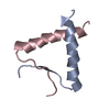
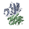
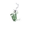
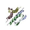
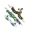
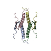
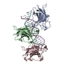
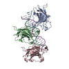
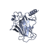
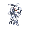
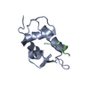
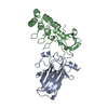
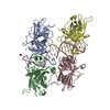
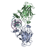
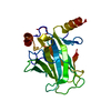
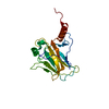
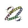
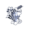
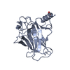
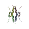
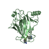
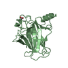
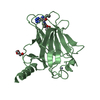
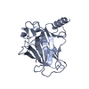
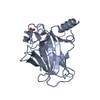
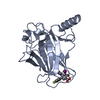
- PDB-2vuk: Structure of the p53 core domain mutant Y220C bound to the stabil... -
+ Open data

- Basic information
| Entry | Database: PDB / ID: 2vuk | ||||||
|---|---|---|---|---|---|---|---|
| Title | Structure of the p53 core domain mutant Y220C bound to the stabilizing small-molecule drug PhiKan083 | ||||||
 Components Components | CELLULAR TUMOR ANTIGEN P53 | ||||||
 Keywords Keywords | TRANSCRIPTION / METAL BINDING / PHOSPHOPROTEIN / UBL CONJUGATION / ACTIVATOR / CELL CYCLE / ACETYLATION / METHYLATION / ZINC / CANCER / NUCLEUS / APOPTOSIS / CYTOPLASM / TUMOR SUPPRESSOR / VIRTUAL SCREENING / SECOND-SITE SUPPRESSOR MUTATION / COVALENT PROTEIN-RNA LINKAGE / SMALL-MOLECULE DRUG / ALTERNATIVE SPLICING / P53 DNA- BINDING DOMAIN / TRANSCRIPTION REGULATION / NUCLEAR PROTEIN / SURFACE CREVICE / DISEASE MUTATION / PROTEIN STABILIZATION / HOST-VIRUS INTERACTION / LI-FRAUMENI SYNDROME / ENDOPLASMIC RETICULUM / METAL-BINDING / ANTI-ONCOGENE / SUPERSTABLE MUTANT / DNA-BINDING PROTEIN / DNA BINDING / DNA-BINDING / POLYMORPHISM / GLYCOPROTEIN | ||||||
| Function / homology |  Function and homology information Function and homology informationnegative regulation of helicase activity / Loss of function of TP53 in cancer due to loss of tetramerization ability / Regulation of TP53 Expression / signal transduction by p53 class mediator / negative regulation of G1 to G0 transition / negative regulation of glucose catabolic process to lactate via pyruvate / Transcriptional activation of cell cycle inhibitor p21 / regulation of intrinsic apoptotic signaling pathway by p53 class mediator / negative regulation of pentose-phosphate shunt / Activation of NOXA and translocation to mitochondria ...negative regulation of helicase activity / Loss of function of TP53 in cancer due to loss of tetramerization ability / Regulation of TP53 Expression / signal transduction by p53 class mediator / negative regulation of G1 to G0 transition / negative regulation of glucose catabolic process to lactate via pyruvate / Transcriptional activation of cell cycle inhibitor p21 / regulation of intrinsic apoptotic signaling pathway by p53 class mediator / negative regulation of pentose-phosphate shunt / Activation of NOXA and translocation to mitochondria / ATP-dependent DNA/DNA annealing activity / regulation of cell cycle G2/M phase transition / oligodendrocyte apoptotic process / negative regulation of miRNA processing / intrinsic apoptotic signaling pathway in response to hypoxia / oxidative stress-induced premature senescence / regulation of tissue remodeling / positive regulation of thymocyte apoptotic process / positive regulation of mitochondrial membrane permeability / germ cell nucleus / regulation of fibroblast apoptotic process / bone marrow development / cellular response to actinomycin D / circadian behavior / histone deacetylase regulator activity / positive regulation of programmed necrotic cell death / : / regulation of mitochondrial membrane permeability involved in apoptotic process / RUNX3 regulates CDKN1A transcription / T cell proliferation involved in immune response / TP53 Regulates Transcription of Death Receptors and Ligands / Activation of PUMA and translocation to mitochondria / TP53 regulates transcription of additional cell cycle genes whose exact role in the p53 pathway remain uncertain / mRNA transcription / negative regulation of glial cell proliferation / negative regulation of neuroblast proliferation / regulation of DNA damage response, signal transduction by p53 class mediator / Regulation of TP53 Activity through Association with Co-factors / Formation of Senescence-Associated Heterochromatin Foci (SAHF) / mitochondrial DNA repair / T cell lineage commitment / thymocyte apoptotic process / ER overload response / TP53 Regulates Transcription of Caspase Activators and Caspases / cardiac septum morphogenesis / B cell lineage commitment / entrainment of circadian clock by photoperiod / negative regulation of DNA replication / Zygotic genome activation (ZGA) / negative regulation of mitophagy / TP53 Regulates Transcription of Genes Involved in Cytochrome C Release / necroptotic process / PI5P Regulates TP53 Acetylation / Association of TriC/CCT with target proteins during biosynthesis / positive regulation of release of cytochrome c from mitochondria / negative regulation of telomere maintenance via telomerase / SUMOylation of transcription factors / TP53 regulates transcription of several additional cell death genes whose specific roles in p53-dependent apoptosis remain uncertain / rRNA transcription / negative regulation of reactive oxygen species metabolic process / TFIID-class transcription factor complex binding / intrinsic apoptotic signaling pathway by p53 class mediator / Transcriptional Regulation by VENTX / cellular response to UV-C / neuroblast proliferation / viral process / intrinsic apoptotic signaling pathway in response to endoplasmic reticulum stress / replicative senescence / positive regulation of RNA polymerase II transcription preinitiation complex assembly / intrinsic apoptotic signaling pathway in response to DNA damage by p53 class mediator / Pyroptosis / general transcription initiation factor binding / chromosome organization / positive regulation of execution phase of apoptosis / hematopoietic stem cell differentiation / embryonic organ development / type II interferon-mediated signaling pathway / TP53 Regulates Transcription of Genes Involved in G1 Cell Cycle Arrest / response to X-ray / hematopoietic progenitor cell differentiation / somitogenesis / negative regulation of stem cell proliferation / core promoter sequence-specific DNA binding / glial cell proliferation / negative regulation of fibroblast proliferation / cis-regulatory region sequence-specific DNA binding / cellular response to glucose starvation / mitophagy / Regulation of TP53 Activity through Acetylation / negative regulation of proteolysis / mitotic G1 DNA damage checkpoint signaling / positive regulation of intrinsic apoptotic signaling pathway / cardiac muscle cell apoptotic process / transcription repressor complex / response to salt stress / 14-3-3 protein binding / gastrulation / positive regulation of cardiac muscle cell apoptotic process / transforming growth factor beta receptor signaling pathway / reactive oxygen species metabolic process Similarity search - Function | ||||||
| Biological species |  HOMO SAPIENS (human) HOMO SAPIENS (human) | ||||||
| Method |  X-RAY DIFFRACTION / SYNCHROTRON / MOLECULAR REPLACEMENT / Resolution: 1.5 Å X-RAY DIFFRACTION / SYNCHROTRON / MOLECULAR REPLACEMENT / Resolution: 1.5 Å | ||||||
 Authors Authors | Joerger, A.C. / Boeckler, F.M. / Fersht, A.R. | ||||||
 Citation Citation | Journal: Proc.Natl.Acad.Sci.USA / Year: 2008 Title: Targeted Rescue of a Destabilized Mutant of P53 by an in Silico Screened Drug. Authors: Boeckler, F.M. / Joerger, A.C. / Jaggi, G. / Rutherford, T.J. / Veprintsev, D.B. / Fersht, A.R. #1: Journal: Proc.Natl.Acad.Sci.USA / Year: 2006Title: Structural Basis for Understanding Oncogenic P53 Mutations and Designing Rescue Drugs Authors: Joerger, A.C. / Ang, H.-C. / Fersht, A.R. | ||||||
| History |
|
- Structure visualization
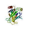
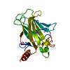
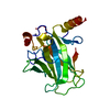
| Structure viewer | Molecule: MolmilJmol/JSmol |
|---|
- Downloads & links
-Download
| PDBx/mmCIF format | 2vuk.cif.gz | 104 KB | Display | PDBx/mmCIF format |
|---|---|---|---|---|
| PDB format | pdb2vuk.ent.gz | 78.7 KB | Display | PDB format |
| PDBx/mmJSON format | 2vuk.json.gz | Tree view | PDBx/mmJSON format | |
| Others |  Other downloads Other downloads |
-Validation report
| Arichive directory | https://data.pdbj.org/pub/pdb/validation_reports/vu/2vukftp://data.pdbj.org/pub/pdb/validation_reports/vu/2vuk | HTTPS FTP |
|---|
-Related structure data
| Related structure data |  2j1xS S: Starting model for refinement |
|---|---|
| Similar structure data |


-Links
 PDBj
PDBj


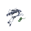
- Assembly
| Deposited unit | 
| ||||||||
|---|---|---|---|---|---|---|---|---|---|
| 1 | 
| ||||||||
| 2 | 
| ||||||||
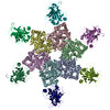
| Unit cell |
|
-Components
| #1: Protein | Mass: 24530.811 Da / Num. of mol.: 2 / Fragment: DNA-BINDING DOMAIN, RESIDUES 94-312 / Mutation: YES Source method: isolated from a genetically manipulated source Source: (gene. exp.) HOMO SAPIENS (human) / Production host:  #2: Chemical |   Mass: 65.409 Da / Num. of mol.: 2 / Source method: obtained synthetically / Formula: Zn Mass: 65.409 Da / Num. of mol.: 2 / Source method: obtained synthetically / Formula: Zn#3: Chemical | ChemComp-P83 / | 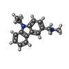  Mass: 238.328 Da / Num. of mol.: 1 / Source method: obtained synthetically / Formula: C16H18N2 Mass: 238.328 Da / Num. of mol.: 1 / Source method: obtained synthetically / Formula: C16H18N2#4: Water | ChemComp-HOH / |  Mass: 18.015 Da / Num. of mol.: 434 / Source method: isolated from a natural source / Formula: H2O Mass: 18.015 Da / Num. of mol.: 434 / Source method: isolated from a natural source / Formula: H2OCompound details | ENGINEERED RESIDUE IN CHAIN A, MET 133 TO LEU ENGINEERED RESIDUE IN CHAIN A, VAL 203 TO ALA ...ENGINEERED | Nonpolymer details | 1-(9-ETHYL-9H-CARBAZOL-3-YL)-N-METHYLMETHANAMINE (P83): STABILIZING SMALL-MOLECULE COMPOUND, ...1-(9-ETHYL-9H-CARBAZOL-3-YL)-N-METHYLMETH | |
|---|
-Experimental details
-Experiment
| Experiment | Method: X-RAY DIFFRACTION / Number of used crystals: 1 |
|---|
- Sample preparation
| Crystal | Density Matthews: 2.5 Å3/Da / Density % sol: 51 % / Description: NONE |
|---|---|
| Crystal grow | Temperature: 294 K / Method: vapor diffusion, sitting drop / pH: 7.2 Details: SITTING DROP VAPOR DIFFUSION AT 21 DEGREE C. PROTEIN SOLUTION: 6 MG/ML PROTEIN IN 25 MM SODIUM PHOSPHATE PH 7.2, 150 MM KCL, 5 MM DTT. RESERVOIR BUFFER: 100 MM HEPES PH 7.2, 19 % PEG 4000, 5 ...Details: SITTING DROP VAPOR DIFFUSION AT 21 DEGREE C. PROTEIN SOLUTION: 6 MG/ML PROTEIN IN 25 MM SODIUM PHOSPHATE PH 7.2, 150 MM KCL, 5 MM DTT. RESERVOIR BUFFER: 100 MM HEPES PH 7.2, 19 % PEG 4000, 5 MM DTT. CRYSTALS WERE SOAKED WITH 10 MM PHIKAN083 IN CRYO BUFFER CONTAINING 100 MM HEPES PH 7.2, 10 MM SODIUM PHOSPHATE PH 7.2, 19 % PEG 4000, 20 % GLYCEROL, 150 MM KCL. |
-Data collection
| Diffraction | Mean temperature: 100 K |
|---|---|
| Diffraction source | Source: SYNCHROTRON / Site: Diamond  / Beamline: I04 / Wavelength: 0.9699 / Beamline: I04 / Wavelength: 0.9699 |
| Detector | Type: ADSC CCD / Detector: CCD / Date: Dec 7, 2007 |
| Radiation | Protocol: SINGLE WAVELENGTH / Monochromatic (M) / Laue (L): M / Scattering type: x-ray |
| Radiation wavelength | Wavelength: 0.9699 Å / Relative weight: 1 |
| Reflection | Resolution: 1.5→65.1 Å / Num. obs: 76025 / % possible obs: 96.6 % / Redundancy: 5.6 % / Biso Wilson estimate: 13.8 Å2 / Rmerge(I) obs: 0.07 / Net I/σ(I): 17.7 |
| Reflection shell | Resolution: 1.5→1.58 Å / Redundancy: 4.6 % / Rmerge(I) obs: 0.23 / Mean I/σ(I) obs: 5.6 / % possible all: 83.4 |
- Processing
| Software |
| ||||||||||||||||||||||||||||||||||||||||||||||||||||||||||||
|---|---|---|---|---|---|---|---|---|---|---|---|---|---|---|---|---|---|---|---|---|---|---|---|---|---|---|---|---|---|---|---|---|---|---|---|---|---|---|---|---|---|---|---|---|---|---|---|---|---|---|---|---|---|---|---|---|---|---|---|---|---|
| Refinement | Method to determine structure: MOLECULAR REPLACEMENT Starting model: PDB ENTRY 2J1X Resolution: 1.5→65.1 Å / Cross valid method: THROUGHOUT
| ||||||||||||||||||||||||||||||||||||||||||||||||||||||||||||
| Displacement parameters | Biso mean: 15.9 Å2 | ||||||||||||||||||||||||||||||||||||||||||||||||||||||||||||
| Refinement step | Cycle: LAST / Resolution: 1.5→65.1 Å
| ||||||||||||||||||||||||||||||||||||||||||||||||||||||||||||
| Refine LS restraints |
|
