 Movie
Movie Controller


+ Open data

- Basic information
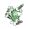
| Entry | Database: PDB / ID: 6si2 | ||||||
|---|---|---|---|---|---|---|---|
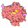
| Title | p53 cancer mutant Y220S | ||||||
 Components Components | Cellular tumor antigen p53 | ||||||
 Keywords Keywords | DNA BINDING PROTEIN / p53 / transcription factor / tumor suppressor / cancer therapy / oncogenic mutant / druggable surface crevice / protein misfolding / mutant p53 rescue / DNA-binding domain | ||||||
| Function / homology |  Function and homology information Function and homology informationnegative regulation of helicase activity / Loss of function of TP53 in cancer due to loss of tetramerization ability / Regulation of TP53 Expression / signal transduction by p53 class mediator / negative regulation of G1 to G0 transition / Transcriptional activation of cell cycle inhibitor p21 / negative regulation of pentose-phosphate shunt / Activation of NOXA and translocation to mitochondria / ATP-dependent DNA/DNA annealing activity / regulation of cell cycle G2/M phase transition ...negative regulation of helicase activity / Loss of function of TP53 in cancer due to loss of tetramerization ability / Regulation of TP53 Expression / signal transduction by p53 class mediator / negative regulation of G1 to G0 transition / Transcriptional activation of cell cycle inhibitor p21 / negative regulation of pentose-phosphate shunt / Activation of NOXA and translocation to mitochondria / ATP-dependent DNA/DNA annealing activity / regulation of cell cycle G2/M phase transition / oligodendrocyte apoptotic process / positive regulation of thymocyte apoptotic process / oxidative stress-induced premature senescence / bone marrow development / circadian behavior / cellular response to actinomycin D / histone deacetylase regulator activity / positive regulation of programmed necrotic cell death / RUNX3 regulates CDKN1A transcription / TP53 Regulates Transcription of Death Receptors and Ligands / Activation of PUMA and translocation to mitochondria / TP53 regulates transcription of additional cell cycle genes whose exact role in the p53 pathway remain uncertain / mRNA transcription / Urea cycle / Regulation of TP53 Activity through Association with Co-factors / ER overload response / hematopoietic stem cell differentiation / Formation of Senescence-Associated Heterochromatin Foci (SAHF) / TP53 Regulates Transcription of Caspase Activators and Caspases / intrinsic apoptotic signaling pathway by p53 class mediator / entrainment of circadian clock by photoperiod / Zygotic genome activation (ZGA) / TP53 Regulates Transcription of Genes Involved in Cytochrome C Release / PI5P Regulates TP53 Acetylation / positive regulation of release of cytochrome c from mitochondria / hematopoietic progenitor cell differentiation / Association of TriC/CCT with target proteins during biosynthesis / negative regulation of telomere maintenance via telomerase / SUMOylation of transcription factors / TP53 regulates transcription of several additional cell death genes whose specific roles in p53-dependent apoptosis remain uncertain / intrinsic apoptotic signaling pathway in response to DNA damage by p53 class mediator / Transcriptional Regulation by VENTX / replicative senescence / TFIID-class transcription factor complex binding / viral process / positive regulation of intrinsic apoptotic signaling pathway / Pyroptosis / determination of adult lifespan / positive regulation of RNA polymerase II transcription preinitiation complex assembly / negative regulation of fibroblast proliferation / general transcription initiation factor binding / intrinsic apoptotic signaling pathway in response to endoplasmic reticulum stress / positive regulation of execution phase of apoptosis / type II interferon-mediated signaling pathway / TP53 Regulates Transcription of Genes Involved in G1 Cell Cycle Arrest / core promoter sequence-specific DNA binding / cellular response to glucose starvation / cis-regulatory region sequence-specific DNA binding / Regulation of TP53 Activity through Acetylation / intrinsic apoptotic signaling pathway / mitotic G1 DNA damage checkpoint signaling / response to gamma radiation / tumor necrosis factor-mediated signaling pathway / 14-3-3 protein binding / MDM2/MDM4 family protein binding / TP53 Regulates Transcription of Genes Involved in G2 Cell Cycle Arrest / transcription repressor complex / protein phosphatase 2A binding / molecular function activator activity / transcription initiation-coupled chromatin remodeling / Regulation of PTEN gene transcription / cellular response to ionizing radiation / cellular response to xenobiotic stimulus / TP53 Regulates Metabolic Genes / DNA damage response, signal transduction by p53 class mediator / TP53 Regulates Transcription of DNA Repair Genes / replication fork / Regulation of NF-kappa B signaling / mRNA 3'-UTR binding / protein tetramerization / promoter-specific chromatin binding / autophagy / Stabilization of p53 / molecular condensate scaffold activity / negative regulation of cell growth / cellular response to gamma radiation / nucleotide-excision repair / G2/M Checkpoints / receptor tyrosine kinase binding / Autodegradation of the E3 ubiquitin ligase COP1 / PML body / positive regulation of miRNA transcription / DNA-binding transcription repressor activity, RNA polymerase II-specific / Oncogene Induced Senescence / PKR-mediated signaling / Regulation of TP53 Activity through Methylation / G2/M DNA damage checkpoint / cellular senescence / DNA Damage/Telomere Stress Induced Senescence / Pre-NOTCH Transcription and Translation Similarity search - Function | ||||||
| Biological species |  Homo sapiens (human) Homo sapiens (human) | ||||||
| Method |  X-RAY DIFFRACTION / SYNCHROTRON / FOURIER SYNTHESIS / Resolution: 1.5 Å X-RAY DIFFRACTION / SYNCHROTRON / FOURIER SYNTHESIS / Resolution: 1.5 Å | ||||||
 Authors Authors | Joerger, A.C. / Bauer, M.R. / Structural Genomics Consortium (SGC) | ||||||
| Funding support |  Germany, 1items Germany, 1items
| ||||||
 Citation Citation | Journal: Acs Chem.Biol. / Year: 2020 Title: Targeting Cavity-Creating p53 Cancer Mutations with Small-Molecule Stabilizers: the Y220X Paradigm. Authors: Bauer, M.R. / Kramer, A. / Settanni, G. / Jones, R.N. / Ni, X. / Khan Tareque, R. / Fersht, A.R. / Spencer, J. / Joerger, A.C. | ||||||
| History |
|
- Structure visualization
| Structure viewer | Molecule: MolmilJmol/JSmol |
|---|
- Downloads & links
-Download
| PDBx/mmCIF format | 6si2.cif.gz | 178.6 KB | Display | PDBx/mmCIF format |
|---|---|---|---|---|
| PDB format | pdb6si2.ent.gz | 140.2 KB | Display | PDB format |
| PDBx/mmJSON format | 6si2.json.gz | Tree view | PDBx/mmJSON format | |
| Others |  Other downloads Other downloads |
-Validation report
| Arichive directory | https://data.pdbj.org/pub/pdb/validation_reports/si/6si2ftp://data.pdbj.org/pub/pdb/validation_reports/si/6si2 | HTTPS FTP |
|---|
-Related structure data
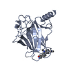
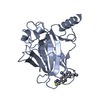
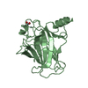
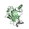
| Related structure data |  6shzC  6si0C  6si1C  6si3C  6si4C  2j1xS S: Starting model for refinement C: citing same article ( |
|---|---|
| Similar structure data |



-Links
 PDBj
PDBj








- Assembly
| Deposited unit | 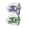
| ||||||||
|---|---|---|---|---|---|---|---|---|---|
| 1 | 
| ||||||||
| 2 | 
| ||||||||
| Unit cell |
|
-Components
| #1: Protein | Mass: 24514.746 Da / Num. of mol.: 2 / Fragment: DNA-binding domain / Mutation: M133L, V203A, Y220S, N239Y, N268D Source method: isolated from a genetically manipulated source Source: (gene. exp.) Homo sapiens (human) / Gene: TP53, P53 / Production host:   Escherichia coli (E. coli) / References: UniProt: P04637 Escherichia coli (E. coli) / References: UniProt: P04637#2: Chemical |   Mass: 65.409 Da / Num. of mol.: 2 / Source method: obtained synthetically / Formula: Zn Mass: 65.409 Da / Num. of mol.: 2 / Source method: obtained synthetically / Formula: Zn#3: Chemical | 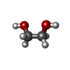 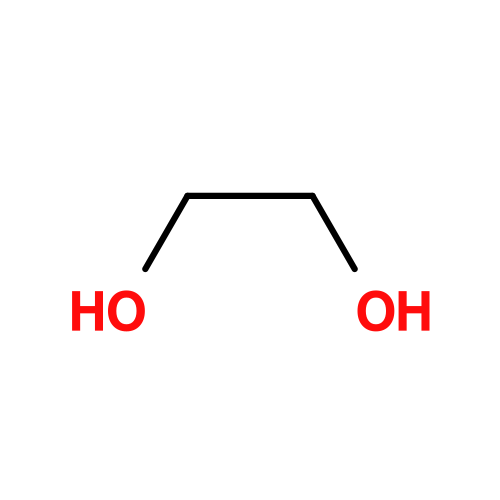 Mass: 62.068 Da / Num. of mol.: 3 / Source method: obtained synthetically / Formula: C2H6O2 Mass: 62.068 Da / Num. of mol.: 3 / Source method: obtained synthetically / Formula: C2H6O2#4: Chemical | ChemComp-GOL / | 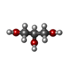 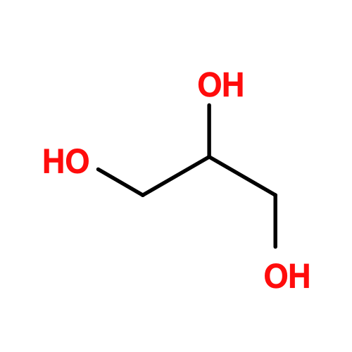 Mass: 92.094 Da / Num. of mol.: 1 / Source method: obtained synthetically / Formula: C3H8O3 Mass: 92.094 Da / Num. of mol.: 1 / Source method: obtained synthetically / Formula: C3H8O3#5: Water | ChemComp-HOH / |  Mass: 18.015 Da / Num. of mol.: 474 / Source method: isolated from a natural source / Formula: H2O Mass: 18.015 Da / Num. of mol.: 474 / Source method: isolated from a natural source / Formula: H2OHas ligand of interest | N | |
|---|
-Experimental details
-Experiment
| Experiment | Method: X-RAY DIFFRACTION / Number of used crystals: 1 |
|---|
- Sample preparation
| Crystal | Density Matthews: 2.5 Å3/Da / Density % sol: 51 % |
|---|---|
| Crystal grow | Temperature: 293 K / Method: vapor diffusion, sitting drop Details: Protein solution: 6 mg/ml protein in 25 mM sodium phosphate, ph 7.2, 150 mm KCl, 5 mm DTT. Reservoir buffer: 100 mm HEPES, pH 7.2, 19% (w/v) polyethylene glycol 4000, 5 mm DTT. |
-Data collection
| Diffraction | Mean temperature: 100 K / Serial crystal experiment: N |
|---|---|
| Diffraction source | Source: SYNCHROTRON / Site: Diamond  / Beamline: I03 / Wavelength: 0.97624 Å / Beamline: I03 / Wavelength: 0.97624 Å |
| Detector | Type: DECTRIS PILATUS 6M / Detector: PIXEL / Date: Aug 8, 2017 |
| Radiation | Protocol: SINGLE WAVELENGTH / Monochromatic (M) / Laue (L): M / Scattering type: x-ray |
| Radiation wavelength | Wavelength: 0.97624 Å / Relative weight: 1 |
| Reflection | Resolution: 1.5→29.6 Å / Num. obs: 78532 / % possible obs: 99.4 % / Redundancy: 4.7 % / CC1/2: 0.999 / Rmerge(I) obs: 0.06 / Net I/σ(I): 14.5 |
| Reflection shell | Resolution: 1.5→1.58 Å / Redundancy: 4.6 % / Rmerge(I) obs: 0.53 / Mean I/σ(I) obs: 2.9 / Num. unique obs: 11369 / CC1/2: 0.875 / % possible all: 99.4 |
- Processing
| Software |
| ||||||||||||||||||||||||||||||||||||||||||||||||||||||||||||||||||||||||||||||||||||||||||||||||||||||||||||||||||||||||||||||||||||||||||||||||||||||||||||||||||||||||||||||||||||
|---|---|---|---|---|---|---|---|---|---|---|---|---|---|---|---|---|---|---|---|---|---|---|---|---|---|---|---|---|---|---|---|---|---|---|---|---|---|---|---|---|---|---|---|---|---|---|---|---|---|---|---|---|---|---|---|---|---|---|---|---|---|---|---|---|---|---|---|---|---|---|---|---|---|---|---|---|---|---|---|---|---|---|---|---|---|---|---|---|---|---|---|---|---|---|---|---|---|---|---|---|---|---|---|---|---|---|---|---|---|---|---|---|---|---|---|---|---|---|---|---|---|---|---|---|---|---|---|---|---|---|---|---|---|---|---|---|---|---|---|---|---|---|---|---|---|---|---|---|---|---|---|---|---|---|---|---|---|---|---|---|---|---|---|---|---|---|---|---|---|---|---|---|---|---|---|---|---|---|---|---|---|
| Refinement | Method to determine structure: FOURIER SYNTHESIS Starting model: 2J1X Resolution: 1.5→29.59 Å / SU ML: 0.12 / Cross valid method: THROUGHOUT / σ(F): 1.34 / Phase error: 17.52
| ||||||||||||||||||||||||||||||||||||||||||||||||||||||||||||||||||||||||||||||||||||||||||||||||||||||||||||||||||||||||||||||||||||||||||||||||||||||||||||||||||||||||||||||||||||
| Solvent computation | Shrinkage radii: 0.9 Å / VDW probe radii: 1.11 Å | ||||||||||||||||||||||||||||||||||||||||||||||||||||||||||||||||||||||||||||||||||||||||||||||||||||||||||||||||||||||||||||||||||||||||||||||||||||||||||||||||||||||||||||||||||||
| Displacement parameters | Biso max: 59.21 Å2 / Biso mean: 22.5537 Å2 / Biso min: 10.4 Å2 | ||||||||||||||||||||||||||||||||||||||||||||||||||||||||||||||||||||||||||||||||||||||||||||||||||||||||||||||||||||||||||||||||||||||||||||||||||||||||||||||||||||||||||||||||||||
| Refinement step | Cycle: final / Resolution: 1.5→29.59 Å
| ||||||||||||||||||||||||||||||||||||||||||||||||||||||||||||||||||||||||||||||||||||||||||||||||||||||||||||||||||||||||||||||||||||||||||||||||||||||||||||||||||||||||||||||||||||
| Refine LS restraints |
| ||||||||||||||||||||||||||||||||||||||||||||||||||||||||||||||||||||||||||||||||||||||||||||||||||||||||||||||||||||||||||||||||||||||||||||||||||||||||||||||||||||||||||||||||||||
| LS refinement shell | Refine-ID: X-RAY DIFFRACTION / Rfactor Rfree error: 0
|
