 Movie
Movie Controller

[English] 日本語
 Yorodumi
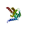
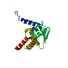
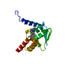
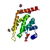
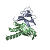
- PDB-2gs0: NMR structure of the complex between the PH domain of the Tfb1 su... -
+ Open data

- Basic information
| Entry | Database: PDB / ID: 2gs0 | ||||||
|---|---|---|---|---|---|---|---|
| Title | NMR structure of the complex between the PH domain of the Tfb1 subunit from TFIIH and the activation domain of p53 | ||||||
 Components Components |
| ||||||
 Keywords Keywords | TRANSCRIPTION / p53 / TFIIH / Tfb1 / activation / PH domain | ||||||
| Function / homology |  Function and homology information Function and homology informationphosphatidylinositol-5-phosphate binding / nucleotide-excision repair factor 3 complex / phosphatidylinositol-3-phosphate binding / transcription factor TFIIH core complex / transcription factor TFIIH holo complex / negative regulation of helicase activity / Loss of function of TP53 in cancer due to loss of tetramerization ability / Regulation of TP53 Expression / signal transduction by p53 class mediator / negative regulation of G1 to G0 transition ...phosphatidylinositol-5-phosphate binding / nucleotide-excision repair factor 3 complex / phosphatidylinositol-3-phosphate binding / transcription factor TFIIH core complex / transcription factor TFIIH holo complex / negative regulation of helicase activity / Loss of function of TP53 in cancer due to loss of tetramerization ability / Regulation of TP53 Expression / signal transduction by p53 class mediator / negative regulation of G1 to G0 transition / negative regulation of glucose catabolic process to lactate via pyruvate / Transcriptional activation of cell cycle inhibitor p21 / regulation of intrinsic apoptotic signaling pathway by p53 class mediator / negative regulation of pentose-phosphate shunt / Activation of NOXA and translocation to mitochondria / ATP-dependent DNA/DNA annealing activity / regulation of cell cycle G2/M phase transition / oligodendrocyte apoptotic process / negative regulation of miRNA processing / intrinsic apoptotic signaling pathway in response to hypoxia / oxidative stress-induced premature senescence / regulation of tissue remodeling / positive regulation of thymocyte apoptotic process / positive regulation of mitochondrial membrane permeability / germ cell nucleus / regulation of fibroblast apoptotic process / bone marrow development / cellular response to actinomycin D / circadian behavior / histone deacetylase regulator activity / positive regulation of programmed necrotic cell death / : / regulation of mitochondrial membrane permeability involved in apoptotic process / RUNX3 regulates CDKN1A transcription / T cell proliferation involved in immune response / TP53 Regulates Transcription of Death Receptors and Ligands / Activation of PUMA and translocation to mitochondria / TP53 regulates transcription of additional cell cycle genes whose exact role in the p53 pathway remain uncertain / mRNA transcription / negative regulation of glial cell proliferation / negative regulation of neuroblast proliferation / regulation of DNA damage response, signal transduction by p53 class mediator / Regulation of TP53 Activity through Association with Co-factors / Formation of Senescence-Associated Heterochromatin Foci (SAHF) / mitochondrial DNA repair / T cell lineage commitment / thymocyte apoptotic process / ER overload response / TP53 Regulates Transcription of Caspase Activators and Caspases / cardiac septum morphogenesis / B cell lineage commitment / RNA Pol II CTD phosphorylation and interaction with CE / Formation of the Early Elongation Complex / mRNA Capping / entrainment of circadian clock by photoperiod / TP53 Regulates Transcription of DNA Repair Genes / RNA Polymerase II Promoter Escape / RNA Polymerase II Transcription Pre-Initiation And Promoter Opening / RNA Polymerase II Transcription Initiation / RNA Polymerase II Transcription Initiation And Promoter Clearance / negative regulation of DNA replication / RNA Polymerase II Pre-transcription Events / Zygotic genome activation (ZGA) / negative regulation of mitophagy / TP53 Regulates Transcription of Genes Involved in Cytochrome C Release / Formation of TC-NER Pre-Incision Complex / necroptotic process / PI5P Regulates TP53 Acetylation / Association of TriC/CCT with target proteins during biosynthesis / positive regulation of release of cytochrome c from mitochondria / negative regulation of telomere maintenance via telomerase / SUMOylation of transcription factors / TP53 regulates transcription of several additional cell death genes whose specific roles in p53-dependent apoptosis remain uncertain / RNA Polymerase I Promoter Escape / rRNA transcription / negative regulation of reactive oxygen species metabolic process / intrinsic apoptotic signaling pathway by p53 class mediator / TFIID-class transcription factor complex binding / Transcriptional Regulation by VENTX / Gap-filling DNA repair synthesis and ligation in TC-NER / cellular response to UV-C / viral process / neuroblast proliferation / intrinsic apoptotic signaling pathway in response to endoplasmic reticulum stress / replicative senescence / intrinsic apoptotic signaling pathway in response to DNA damage by p53 class mediator / Dual incision in TC-NER / Pyroptosis / positive regulation of RNA polymerase II transcription preinitiation complex assembly / chromosome organization / general transcription initiation factor binding / positive regulation of execution phase of apoptosis / hematopoietic stem cell differentiation / embryonic organ development / type II interferon-mediated signaling pathway / TP53 Regulates Transcription of Genes Involved in G1 Cell Cycle Arrest / response to X-ray / hematopoietic progenitor cell differentiation / somitogenesis / negative regulation of stem cell proliferation Similarity search - Function | ||||||
| Biological species |   Homo sapiens (human) Homo sapiens (human) | ||||||
| Method | SOLUTION NMR / Simulated annealing, with a combination of torsion angle, Cartesian dynamics. | ||||||
 Authors Authors | Di Lello, P. / Jones, T.N. / Nguyen, B.D. / Legault, P. / Omichinski, J.G. | ||||||
 Citation Citation | Journal: Mol.Cell / Year: 2006 Title: Structure of the Tfb1/p53 complex: Insights into the interaction between the p62/Tfb1 subunit of TFIIH and the activation domain of p53. Authors: Di Lello, P. / Jenkins, L.M. / Jones, T.N. / Nguyen, B.D. / Hara, T. / Yamaguchi, H. / Dikeakos, J.D. / Appella, E. / Legault, P. / Omichinski, J.G. | ||||||
| History |
|
- Structure visualization
| Structure viewer | Molecule: MolmilJmol/JSmol |
|---|
- Downloads & links
-Download
| PDBx/mmCIF format | 2gs0.cif.gz | 793.9 KB | Display | PDBx/mmCIF format |
|---|---|---|---|---|
| PDB format | pdb2gs0.ent.gz | 662.8 KB | Display | PDB format |
| PDBx/mmJSON format | 2gs0.json.gz | Tree view | PDBx/mmJSON format | |
| Others |  Other downloads Other downloads |
-Validation report
| Arichive directory | https://data.pdbj.org/pub/pdb/validation_reports/gs/2gs0ftp://data.pdbj.org/pub/pdb/validation_reports/gs/2gs0 | HTTPS FTP |
|---|
-Related structure data
| Similar structure data |
|---|

-Links
 PDBj
PDBj


- Assembly
| Deposited unit | 
| |||||||||
|---|---|---|---|---|---|---|---|---|---|---|
| 1 |
| |||||||||
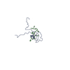
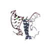
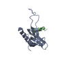
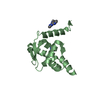
| NMR ensembles |
|
-Components
| #1: Protein | Mass: 12903.701 Da / Num. of mol.: 1 / Mutation: M1P Source method: isolated from a genetically manipulated source Source: (gene. exp.) Gene: TFB1 / Plasmid: pGEX-2T / Production host:  |
|---|---|
| #2: Protein | Mass: 6018.665 Da / Num. of mol.: 1 Source method: isolated from a genetically manipulated source Source: (gene. exp.) Homo sapiens (human) / Gene: TP53 / Plasmid: pGEX-2TK / Production host: |
-Experimental details
-Experiment
| Experiment | Method: SOLUTION NMR | ||||||||||||||||||||||||||||||||||||||||||||
|---|---|---|---|---|---|---|---|---|---|---|---|---|---|---|---|---|---|---|---|---|---|---|---|---|---|---|---|---|---|---|---|---|---|---|---|---|---|---|---|---|---|---|---|---|---|
| NMR experiment |
| ||||||||||||||||||||||||||||||||||||||||||||
| NMR details | Text: The structure was determined using triple-resonance NMR spectroscopy. |
 HSQC
HSQC- Sample preparation
| Details |
| |||||||||||||||
|---|---|---|---|---|---|---|---|---|---|---|---|---|---|---|---|---|
| Sample conditions | Ionic strength: 20 mM sodium phosphate / pH: 6.5 / Pressure: ambient / Temperature: 300 K |
-NMR measurement
| Radiation | Protocol: SINGLE WAVELENGTH / Monochromatic (M) / Laue (L): M | ||||||||||||||||||||
|---|---|---|---|---|---|---|---|---|---|---|---|---|---|---|---|---|---|---|---|---|---|
| Radiation wavelength | Relative weight: 1 | ||||||||||||||||||||
| NMR spectrometer |
|
- Processing
| NMR software |
| ||||||||||||||||||||||||
|---|---|---|---|---|---|---|---|---|---|---|---|---|---|---|---|---|---|---|---|---|---|---|---|---|---|
| Refinement | Method: Simulated annealing, with a combination of torsion angle, Cartesian dynamics. Software ordinal: 1 Details: The three-dimensional structures of the complex Tfb1/p53 were determined using a set of 1720 NOE-derived distance restraints, 138 backbone dihedral angle (phi and psi) restraints and 26 ...Details: The three-dimensional structures of the complex Tfb1/p53 were determined using a set of 1720 NOE-derived distance restraints, 138 backbone dihedral angle (phi and psi) restraints and 26 distance restraints from hydrogen bonds. Because of the absence of medium-range, long-range and intermolecular NOEs involving residues 20-44 and 59-73 of p53 (chain B), these amino acids were not included in the calculations. | ||||||||||||||||||||||||
| NMR representative | Selection criteria: lowest energy | ||||||||||||||||||||||||
| NMR ensemble | Conformer selection criteria: structures with the lowest energy Conformers calculated total number: 67 / Conformers submitted total number: 20 |
