 Movie
Movie Controller

[English] 日本語
 Yorodumi
- PDB-4g7x: Crystal structure of a complex between the CTXphi pIII N-terminal... -
+ Open data

- Basic information
| Entry | Database: PDB / ID: 4g7x | ||||||
|---|---|---|---|---|---|---|---|
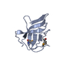
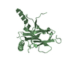
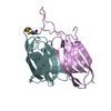
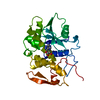
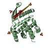
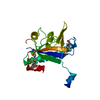
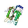
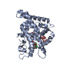
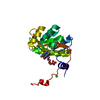
| Title | Crystal structure of a complex between the CTXphi pIII N-terminal domain and the Vibrio cholerae TolA C-terminal domain | ||||||
 Components Components |
| ||||||
 Keywords Keywords | PROTEIN BINDING/PROTEIN BINDING / Membrane / PROTEIN BINDING-PROTEIN BINDING complex | ||||||
| Function / homology |  Function and homology information Function and homology informationbacteriocin transport / toxin transmembrane transporter activity / membrane => GO:0016020 / membrane Similarity search - Function | ||||||
| Biological species |   Vibrio cholerae (bacteria) Vibrio cholerae (bacteria) | ||||||
| Method |  X-RAY DIFFRACTION / SYNCHROTRON / MOLECULAR REPLACEMENT / Resolution: 1.44 Å X-RAY DIFFRACTION / SYNCHROTRON / MOLECULAR REPLACEMENT / Resolution: 1.44 Å | ||||||
 Authors Authors | Kolappan, S. / Ford, C.G. / Craig, L. | ||||||
 Citation Citation | Journal: J.Biol.Chem. / Year: 2012 Title: Crystal Structures of a CTX{varphi} pIII Domain Unbound and in Complex with a Vibrio cholerae TolA Domain Reveal Novel Interaction Interfaces. Authors: Ford, C.G. / Kolappan, S. / Phan, H.T. / Waldor, M.K. / Winther-Larsen, H.C. / Craig, L. | ||||||
| History |
|
- Structure visualization
| Structure viewer | Molecule: MolmilJmol/JSmol |
|---|
- Downloads & links
-Download
| PDBx/mmCIF format | 4g7x.cif.gz | 58.9 KB | Display | PDBx/mmCIF format |
|---|---|---|---|---|
| PDB format | pdb4g7x.ent.gz | 41.9 KB | Display | PDB format |
| PDBx/mmJSON format | 4g7x.json.gz | Tree view | PDBx/mmJSON format | |
| Others |  Other downloads Other downloads |
-Validation report
| Arichive directory | https://data.pdbj.org/pub/pdb/validation_reports/g7/4g7xftp://data.pdbj.org/pub/pdb/validation_reports/g7/4g7x | HTTPS FTP |
|---|
-Related structure data


-Links
 PDBj
PDBj- Assembly
| Deposited unit | 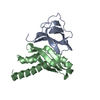
| ||||||||
|---|---|---|---|---|---|---|---|---|---|
| 1 |
| ||||||||
| Unit cell |
|
-Components
| #1: Protein | Mass: 11509.833 Da / Num. of mol.: 1 / Fragment: N-TERMINAL DOMAIN Source method: isolated from a genetically manipulated source Source: (gene. exp.) Vibrio cholerae (bacteria) / Strain: CIRS101 / Gene: orfU, VCH_002198 / Production host: |
|---|---|
| #2: Protein | Mass: 15072.992 Da / Num. of mol.: 1 / Fragment: C-TERMINAL DOMAIN Source method: isolated from a genetically manipulated source Source: (gene. exp.) Vibrio cholerae (bacteria) / Strain: ATCC 39541 / Ogawa 395 / O395 / Gene: tolA, VC0395_A1430, VC395_1952 / Production host: |
| #3: Water | ChemComp-HOH /  Mass: 18.015 Da / Num. of mol.: 197 / Source method: isolated from a natural source / Formula: H2O Mass: 18.015 Da / Num. of mol.: 197 / Source method: isolated from a natural source / Formula: H2O |
| Has protein modification | Y |
-Experimental details
-Experiment
| Experiment | Method: X-RAY DIFFRACTION / Number of used crystals: 1 |
|---|
- Sample preparation
| Crystal | Density Matthews: 1.91 Å3/Da / Density % sol: 35.73 % |
|---|---|
| Crystal grow | Temperature: 293 K / Method: vapor diffusion, hanging drop / pH: 6 Details: 25 % PEG 6000, 100 mM MES, pH 6.0, VAPOR DIFFUSION, HANGING DROP, temperature 293K |
-Data collection
| Diffraction | Mean temperature: 100 K |
|---|---|
| Diffraction source | Source: SYNCHROTRON / Site: SSRL  / Beamline: BL9-2 / Wavelength: 1 Å / Beamline: BL9-2 / Wavelength: 1 Å |
| Detector | Type: MARMOSAIC 325 mm CCD / Detector: CCD / Date: Dec 13, 2011 / Details: mirrors |
| Radiation | Monochromator: Double crystal monochromator, Si(111) / Protocol: SINGLE WAVELENGTH / Monochromatic (M) / Laue (L): M / Scattering type: x-ray |
| Radiation wavelength | Wavelength: 1 Å / Relative weight: 1 |
| Reflection | Resolution: 1.44→39.9 Å / Num. all: 37355 / Num. obs: 35426 / % possible obs: 99.6 % / Observed criterion σ(F): 2 / Observed criterion σ(I): 4.4 / Redundancy: 6.9 % / Biso Wilson estimate: 19.8 Å2 / Rsym value: 0.042 / Net I/σ(I): 27.3 |
| Reflection shell | Resolution: 1.44→1.48 Å / Redundancy: 4.6 % / Mean I/σ(I) obs: 4.4 / Num. unique all: 257179 / Rsym value: 0.329 / % possible all: 95.5 |
- Processing
| Software |
| |||||||||||||||||||||||||||||||||||||||||||||
|---|---|---|---|---|---|---|---|---|---|---|---|---|---|---|---|---|---|---|---|---|---|---|---|---|---|---|---|---|---|---|---|---|---|---|---|---|---|---|---|---|---|---|---|---|---|---|
| Refinement | Method to determine structure: MOLECULAR REPLACEMENT / Resolution: 1.44→39.9 Å / Cor.coef. Fo:Fc: 0.948 / Cor.coef. Fo:Fc free: 0.945 / SU B: 1.114 / SU ML: 0.045 / Cross valid method: THROUGHOUT / σ(F): 2 / ESU R: 0.078 / ESU R Free: 0.073 / Stereochemistry target values: MAXIMUM LIKELIHOOD / Details: HYDROGENS HAVE BEEN USED IF PRESENT IN THE INPUT
| |||||||||||||||||||||||||||||||||||||||||||||
| Solvent computation | Ion probe radii: 0.8 Å / Shrinkage radii: 0.8 Å / VDW probe radii: 1.2 Å / Solvent model: MASK | |||||||||||||||||||||||||||||||||||||||||||||
| Displacement parameters | Biso mean: 14.936 Å2
| |||||||||||||||||||||||||||||||||||||||||||||
| Refinement step | Cycle: LAST / Resolution: 1.44→39.9 Å
| |||||||||||||||||||||||||||||||||||||||||||||
| Refine LS restraints |
| |||||||||||||||||||||||||||||||||||||||||||||
| LS refinement shell | Resolution: 1.44→1.482 Å / Total num. of bins used: 20
|
