 Movie
Movie Controller

[English] 日本語
 Yorodumi
- PDB-5cuz: Crystal structure of SeMet-substituted N-terminal truncated human... -
+ Open data

- Basic information
| Entry | Database: PDB / ID: 5cuz | ||||||
|---|---|---|---|---|---|---|---|
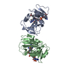
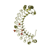
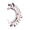
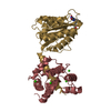
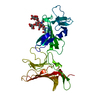
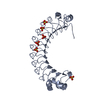
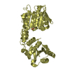
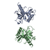
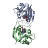
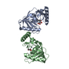
| Title | Crystal structure of SeMet-substituted N-terminal truncated human B12-chaperone CblD (108-296) | ||||||
 Components Components | Methylmalonic aciduria and homocystinuria type D protein, mitochondrial | ||||||
 Keywords Keywords | CHAPERONE / Vitamin B12 / Nitro-FMN-reductase | ||||||
| Function / homology |  Function and homology information Function and homology informationDefective MMADHC causes MMAHCD / Cobalamin (Cbl) metabolism / cobalamin metabolic process / molecular carrier activity / mitochondrion / cytoplasm / cytosol Similarity search - Function | ||||||
| Biological species |  Homo sapiens (human) Homo sapiens (human) | ||||||
| Method |  X-RAY DIFFRACTION / SYNCHROTRON / SAD / Resolution: 2.31 Å X-RAY DIFFRACTION / SYNCHROTRON / SAD / Resolution: 2.31 Å | ||||||
 Authors Authors | Yamada, K. / Gherasim, C. / Banerjee, R. / Koutmos, M. | ||||||
| Funding support |  United States, 1items United States, 1items
| ||||||
 Citation Citation | Journal: J.Biol.Chem. / Year: 2015 Title: Structure of Human B12 Trafficking Protein CblD Reveals Molecular Mimicry and Identifies a New Subfamily of Nitro-FMN Reductases. Authors: Yamada, K. / Gherasim, C. / Banerjee, R. / Koutmos, M. | ||||||
| History |
|
- Structure visualization
| Structure viewer | Molecule: MolmilJmol/JSmol |
|---|
- Downloads & links
-Download
| PDBx/mmCIF format | 5cuz.cif.gz | 70.9 KB | Display | PDBx/mmCIF format |
|---|---|---|---|---|
| PDB format | pdb5cuz.ent.gz | 53.9 KB | Display | PDB format |
| PDBx/mmJSON format | 5cuz.json.gz | Tree view | PDBx/mmJSON format | |
| Others |  Other downloads Other downloads |
-Validation report
| Arichive directory | https://data.pdbj.org/pub/pdb/validation_reports/cu/5cuzftp://data.pdbj.org/pub/pdb/validation_reports/cu/5cuz | HTTPS FTP |
|---|
-Related structure data

-Links
 PDBj
PDBj
- Assembly
| Deposited unit | 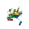
| ||||||||
|---|---|---|---|---|---|---|---|---|---|
| 1 | 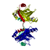
| ||||||||
| Unit cell |
|
-Components
| #1: Protein | Mass: 22074.982 Da / Num. of mol.: 1 / Fragment: UNP residues 108-296 Source method: isolated from a genetically manipulated source Source: (gene. exp.) Homo sapiens (human) / Gene: MMADHC, C2orf25, CL25022, HSPC161, My011 / Plasmid: pET28b(+) / Production host:  |
|---|---|
| #2: Water | ChemComp-HOH /  Mass: 18.015 Da / Num. of mol.: 21 / Source method: isolated from a natural source / Formula: H2O Mass: 18.015 Da / Num. of mol.: 21 / Source method: isolated from a natural source / Formula: H2O |
| Has protein modification | Y |
-Experimental details
-Experiment
| Experiment | Method: X-RAY DIFFRACTION |
|---|
- Sample preparation
| Crystal | Density Matthews: 1.8 Å3/Da / Density % sol: 32.2 % |
|---|---|
| Crystal grow | Temperature: 293 K / Method: vapor diffusion, sitting drop / pH: 7.5 Details: 20 % PEG3350, 0.1M Tris-HCl, pH 7.5, 0.185 M MgCl2, 0.12 M NaF |
-Data collection
| Diffraction | Mean temperature: 100 K |
|---|---|
| Diffraction source | Source: SYNCHROTRON / Site: APS / Beamline: 23-ID-B / Wavelength: 0.979475 Å |
| Detector | Type: MARMOSAIC 300 mm CCD / Detector: CCD / Date: Mar 23, 2013 Details: K-B pair of biomorph mirrors for vertical and horizontal focusing |
| Radiation | Monochromator: double crystal monochromator / Protocol: SINGLE WAVELENGTH / Monochromatic (M) / Laue (L): M / Scattering type: x-ray |
| Radiation wavelength | Wavelength: 0.979475 Å / Relative weight: 1 |
| Reflection | Resolution: 2.31→50 Å / Num. obs: 6667 / % possible obs: 94.1 % / Redundancy: 7 % / Rmerge(I) obs: 0.098 / Net I/σ(I): 14.5 |
| Reflection shell | Resolution: 2.31→2.39 Å / Redundancy: 3.5 % / Rmerge(I) obs: 0.359 / Mean I/σ(I) obs: 2.5 / % possible all: 70.4 |
- Processing
| Software |
| ||||||||||||||||||||||||||||||||||||||||||||||||||||||||||||||||||||||||||||||||||||||||||||||||||||||||||||||||||||||||||||||||||||||||||||||||||||||||||||||||||||||||||||||||||||||
|---|---|---|---|---|---|---|---|---|---|---|---|---|---|---|---|---|---|---|---|---|---|---|---|---|---|---|---|---|---|---|---|---|---|---|---|---|---|---|---|---|---|---|---|---|---|---|---|---|---|---|---|---|---|---|---|---|---|---|---|---|---|---|---|---|---|---|---|---|---|---|---|---|---|---|---|---|---|---|---|---|---|---|---|---|---|---|---|---|---|---|---|---|---|---|---|---|---|---|---|---|---|---|---|---|---|---|---|---|---|---|---|---|---|---|---|---|---|---|---|---|---|---|---|---|---|---|---|---|---|---|---|---|---|---|---|---|---|---|---|---|---|---|---|---|---|---|---|---|---|---|---|---|---|---|---|---|---|---|---|---|---|---|---|---|---|---|---|---|---|---|---|---|---|---|---|---|---|---|---|---|---|---|---|
| Refinement | Method to determine structure: SAD / Resolution: 2.31→48.71 Å / Cor.coef. Fo:Fc: 0.942 / Cor.coef. Fo:Fc free: 0.901 / SU B: 8.472 / SU ML: 0.2 / Cross valid method: THROUGHOUT / ESU R: 0.521 / ESU R Free: 0.284 / Stereochemistry target values: MAXIMUM LIKELIHOOD / Details: HYDROGENS HAVE BEEN ADDED IN THE RIDING POSITIONS
| ||||||||||||||||||||||||||||||||||||||||||||||||||||||||||||||||||||||||||||||||||||||||||||||||||||||||||||||||||||||||||||||||||||||||||||||||||||||||||||||||||||||||||||||||||||||
| Solvent computation | Ion probe radii: 0.8 Å / Shrinkage radii: 0.8 Å / VDW probe radii: 1.2 Å / Solvent model: BABINET MODEL WITH MASK | ||||||||||||||||||||||||||||||||||||||||||||||||||||||||||||||||||||||||||||||||||||||||||||||||||||||||||||||||||||||||||||||||||||||||||||||||||||||||||||||||||||||||||||||||||||||
| Displacement parameters | Biso mean: 45.151 Å2
| ||||||||||||||||||||||||||||||||||||||||||||||||||||||||||||||||||||||||||||||||||||||||||||||||||||||||||||||||||||||||||||||||||||||||||||||||||||||||||||||||||||||||||||||||||||||
| Refinement step | Cycle: 1 / Resolution: 2.31→48.71 Å
| ||||||||||||||||||||||||||||||||||||||||||||||||||||||||||||||||||||||||||||||||||||||||||||||||||||||||||||||||||||||||||||||||||||||||||||||||||||||||||||||||||||||||||||||||||||||
| Refine LS restraints |
|