 Movie
Movie Controller

[English] 日本語
 Yorodumi
- PDB-2h1l: The Structure of the Oncoprotein SV40 Large T Antigen and p53 Tum... -
+ Open data

- Basic information
| Entry | Database: PDB / ID: 2h1l | ||||||
|---|---|---|---|---|---|---|---|
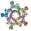
| Title | The Structure of the Oncoprotein SV40 Large T Antigen and p53 Tumor Suppressor Complex | ||||||
 Components Components |
| ||||||
 Keywords Keywords | VIRAL PROTEIN / p53 Loop-3 conformation change | ||||||
| Function / homology |  Function and homology information Function and homology informationsymbiont-mediated suppression of host JAK-STAT cascade via inhibition of JAK1 activity / negative regulation of helicase activity / Loss of function of TP53 in cancer due to loss of tetramerization ability / Regulation of TP53 Expression / signal transduction by p53 class mediator / negative regulation of G1 to G0 transition / negative regulation of glucose catabolic process to lactate via pyruvate / Transcriptional activation of cell cycle inhibitor p21 / regulation of intrinsic apoptotic signaling pathway by p53 class mediator / negative regulation of pentose-phosphate shunt ...symbiont-mediated suppression of host JAK-STAT cascade via inhibition of JAK1 activity / negative regulation of helicase activity / Loss of function of TP53 in cancer due to loss of tetramerization ability / Regulation of TP53 Expression / signal transduction by p53 class mediator / negative regulation of G1 to G0 transition / negative regulation of glucose catabolic process to lactate via pyruvate / Transcriptional activation of cell cycle inhibitor p21 / regulation of intrinsic apoptotic signaling pathway by p53 class mediator / negative regulation of pentose-phosphate shunt / bidirectional double-stranded viral DNA replication / Activation of NOXA and translocation to mitochondria / ATP-dependent DNA/DNA annealing activity / regulation of cell cycle G2/M phase transition / oligodendrocyte apoptotic process / negative regulation of miRNA processing / intrinsic apoptotic signaling pathway in response to hypoxia / oxidative stress-induced premature senescence / regulation of tissue remodeling / positive regulation of thymocyte apoptotic process / viral DNA genome replication / positive regulation of mitochondrial membrane permeability / germ cell nucleus / regulation of fibroblast apoptotic process / bone marrow development / circadian behavior / histone deacetylase regulator activity / positive regulation of programmed necrotic cell death / cellular response to actinomycin D / : / regulation of mitochondrial membrane permeability involved in apoptotic process / RUNX3 regulates CDKN1A transcription / T cell proliferation involved in immune response / TP53 Regulates Transcription of Death Receptors and Ligands / Activation of PUMA and translocation to mitochondria / TP53 regulates transcription of additional cell cycle genes whose exact role in the p53 pathway remain uncertain / mRNA transcription / negative regulation of glial cell proliferation / regulation of DNA damage response, signal transduction by p53 class mediator / Regulation of TP53 Activity through Association with Co-factors / negative regulation of neuroblast proliferation / Formation of Senescence-Associated Heterochromatin Foci (SAHF) / mitochondrial DNA repair / T cell lineage commitment / thymocyte apoptotic process / ER overload response / TP53 Regulates Transcription of Caspase Activators and Caspases / cardiac septum morphogenesis / B cell lineage commitment / entrainment of circadian clock by photoperiod / negative regulation of DNA replication / Zygotic genome activation (ZGA) / negative regulation of mitophagy / symbiont-mediated perturbation of host cell cycle G1/S transition checkpoint / TP53 Regulates Transcription of Genes Involved in Cytochrome C Release / PI5P Regulates TP53 Acetylation / necroptotic process / negative regulation of telomere maintenance via telomerase / Association of TriC/CCT with target proteins during biosynthesis / positive regulation of release of cytochrome c from mitochondria / SUMOylation of transcription factors / TP53 regulates transcription of several additional cell death genes whose specific roles in p53-dependent apoptosis remain uncertain / 3'-5' DNA helicase activity / DNA 3'-5' helicase / rRNA transcription / negative regulation of reactive oxygen species metabolic process / TFIID-class transcription factor complex binding / intrinsic apoptotic signaling pathway by p53 class mediator / Transcriptional Regulation by VENTX / cellular response to UV-C / viral process / neuroblast proliferation / DNA replication origin binding / intrinsic apoptotic signaling pathway in response to endoplasmic reticulum stress / replicative senescence / positive regulation of RNA polymerase II transcription preinitiation complex assembly / Pyroptosis / intrinsic apoptotic signaling pathway in response to DNA damage by p53 class mediator / general transcription initiation factor binding / chromosome organization / positive regulation of execution phase of apoptosis / hematopoietic stem cell differentiation / type II interferon-mediated signaling pathway / embryonic organ development / response to X-ray / TP53 Regulates Transcription of Genes Involved in G1 Cell Cycle Arrest / hematopoietic progenitor cell differentiation / somitogenesis / negative regulation of stem cell proliferation / core promoter sequence-specific DNA binding / glial cell proliferation / cellular response to glucose starvation / cis-regulatory region sequence-specific DNA binding / negative regulation of fibroblast proliferation / mitophagy / Regulation of TP53 Activity through Acetylation / negative regulation of proteolysis / mitotic G1 DNA damage checkpoint signaling / response to salt stress / positive regulation of intrinsic apoptotic signaling pathway Similarity search - Function | ||||||
| Biological species |  Simian virus 40 Simian virus 40 Homo sapiens (human) Homo sapiens (human) | ||||||
| Method |  X-RAY DIFFRACTION / SYNCHROTRON / MOLECULAR REPLACEMENT / Resolution: 3.16 Å X-RAY DIFFRACTION / SYNCHROTRON / MOLECULAR REPLACEMENT / Resolution: 3.16 Å | ||||||
 Authors Authors | Lilyestrom, W. / Klein, M.G. / Chen, X.S. | ||||||
 Citation Citation | Journal: Genes Dev. / Year: 2006 Title: Crystal structure of SV40 large T-antigen bound to p53: interplay between a viral oncoprotein and a cellular tumor suppressor. Authors: Lilyestrom, W. / Klein, M.G. / Zhang, R. / Joachimiak, A. / Chen, X.S. | ||||||
| History |
|
- Structure visualization
| Structure viewer | Molecule: MolmilJmol/JSmol |
|---|
- Downloads & links
-Download
| PDBx/mmCIF format | 2h1l.cif.gz | 1.3 MB | Display | PDBx/mmCIF format |
|---|---|---|---|---|
| PDB format | pdb2h1l.ent.gz | 1.1 MB | Display | PDB format |
| PDBx/mmJSON format | 2h1l.json.gz | Tree view | PDBx/mmJSON format | |
| Others |  Other downloads Other downloads |
-Validation report
| Arichive directory | https://data.pdbj.org/pub/pdb/validation_reports/h1/2h1lftp://data.pdbj.org/pub/pdb/validation_reports/h1/2h1l | HTTPS FTP |
|---|
-Related structure data
| Related structure data | 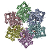 1svmS S: Starting model for refinement |
|---|---|
| Similar structure data |


-Links
 PDBj
PDBj


- Assembly
| Deposited unit | 
| ||||||||
|---|---|---|---|---|---|---|---|---|---|
| 1 | 
| ||||||||
| 2 | 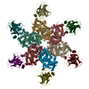
| ||||||||
| Unit cell |
| ||||||||
| Details | The biological assembly is a single hexamer containing six LT and six p53 molecules. There are two such hexamers in the asu. The first contains chains A-F and M-R. The is composed of chains G-L and S-X. |
-Components
| #1: Protein | Mass: 42645.426 Da / Num. of mol.: 12 / Fragment: Helicase Domain, residues 260-627 Source method: isolated from a genetically manipulated source Source: (gene. exp.) Simian virus 40 / Genus: Polyomavirus / Gene: Gene A / Plasmid: pGEX-KG / Production host:  #2: Protein | Mass: 22802.936 Da / Num. of mol.: 12 / Fragment: DNA Binding Domain, residues 92-292 Source method: isolated from a genetically manipulated source Source: (gene. exp.) Homo sapiens (human) / Gene: p53 / Plasmid: pGEX-KG / Production host: #3: Chemical | ChemComp-ZN /   Mass: 65.409 Da / Num. of mol.: 24 / Source method: obtained synthetically / Formula: Zn Mass: 65.409 Da / Num. of mol.: 24 / Source method: obtained synthetically / Formula: ZnHas protein modification | Y | |
|---|
-Experimental details
-Experiment
| Experiment | Method: X-RAY DIFFRACTION / Number of used crystals: 1 |
|---|
- Sample preparation
| Crystal | Density Matthews: 3.88 Å3/Da / Density % sol: 68.3 % |
|---|---|
| Crystal grow | Temperature: 298 K / Method: vapor diffusion / pH: 6.5 Details: 5.4% PEG 4000, 73mM Mops Buffer, 22mM Lithium Sulfate, pH 6.5, VAPOR DIFFUSION, temperature 298K |
-Data collection
| Diffraction | Mean temperature: 200 K |
|---|---|
| Diffraction source | Source: SYNCHROTRON / Site: APS  / Beamline: 19-ID / Wavelength: 0.98 Å / Beamline: 19-ID / Wavelength: 0.98 Å |
| Detector | Type: ADSC QUANTUM 315 / Detector: CCD / Date: Jul 12, 2005 |
| Radiation | Protocol: SINGLE WAVELENGTH / Monochromatic (M) / Laue (L): M / Scattering type: x-ray |
| Radiation wavelength | Wavelength: 0.98 Å / Relative weight: 1 |
| Reflection | Resolution: 3.16→20 Å / Num. obs: 204669 / % possible obs: 97.5 % / Observed criterion σ(F): 2 / Observed criterion σ(I): 2 / Redundancy: 3.7 % / Rsym value: 0.069 / Net I/σ(I): 30.3 |
| Reflection shell | Resolution: 3.16→3.27 Å / Redundancy: 3.2 % / Mean I/σ(I) obs: 4.5 / Num. unique all: 204669 / Rsym value: 0.398 / % possible all: 94.3 |
- Processing
| Software |
| |||||||||||||||||||||||||
|---|---|---|---|---|---|---|---|---|---|---|---|---|---|---|---|---|---|---|---|---|---|---|---|---|---|---|
| Refinement | Method to determine structure: MOLECULAR REPLACEMENT Starting model: PDB ENTRY 1SVM Resolution: 3.16→20 Å / σ(F): 2 / σ(I): 2 / Stereochemistry target values: Engh & Huber
| |||||||||||||||||||||||||
| Displacement parameters | Biso mean: 75.3 Å2 | |||||||||||||||||||||||||
| Refinement step | Cycle: LAST / Resolution: 3.16→20 Å
| |||||||||||||||||||||||||
| Refine LS restraints |
| |||||||||||||||||||||||||
| LS refinement shell | Resolution: 3.16→3.27 Å / Num. reflection obs: 19180 | |||||||||||||||||||||||||
| Xplor file |
|
