 Movie
Movie Controller


+ Open data

- Basic information
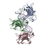
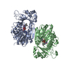
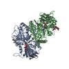
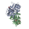
| Entry | Database: PDB / ID: 1tup | ||||||
|---|---|---|---|---|---|---|---|
| Title | TUMOR SUPPRESSOR P53 COMPLEXED WITH DNA | ||||||
 Components Components |
| ||||||
 Keywords Keywords | ANTITUMOR PROTEIN/DNA / ANTIGEN P53 / ANTITUMOR PROTEIN-DNA COMPLEX | ||||||
| Function / homology |  Function and homology information Function and homology informationnegative regulation of helicase activity / Loss of function of TP53 in cancer due to loss of tetramerization ability / Regulation of TP53 Expression / signal transduction by p53 class mediator / negative regulation of G1 to G0 transition / negative regulation of glucose catabolic process to lactate via pyruvate / Transcriptional activation of cell cycle inhibitor p21 / regulation of intrinsic apoptotic signaling pathway by p53 class mediator / negative regulation of pentose-phosphate shunt / Activation of NOXA and translocation to mitochondria ...negative regulation of helicase activity / Loss of function of TP53 in cancer due to loss of tetramerization ability / Regulation of TP53 Expression / signal transduction by p53 class mediator / negative regulation of G1 to G0 transition / negative regulation of glucose catabolic process to lactate via pyruvate / Transcriptional activation of cell cycle inhibitor p21 / regulation of intrinsic apoptotic signaling pathway by p53 class mediator / negative regulation of pentose-phosphate shunt / Activation of NOXA and translocation to mitochondria / ATP-dependent DNA/DNA annealing activity / regulation of cell cycle G2/M phase transition / oligodendrocyte apoptotic process / negative regulation of miRNA processing / intrinsic apoptotic signaling pathway in response to hypoxia / oxidative stress-induced premature senescence / regulation of tissue remodeling / positive regulation of thymocyte apoptotic process / positive regulation of mitochondrial membrane permeability / germ cell nucleus / regulation of fibroblast apoptotic process / bone marrow development / cellular response to actinomycin D / circadian behavior / histone deacetylase regulator activity / positive regulation of programmed necrotic cell death / : / regulation of mitochondrial membrane permeability involved in apoptotic process / RUNX3 regulates CDKN1A transcription / T cell proliferation involved in immune response / TP53 Regulates Transcription of Death Receptors and Ligands / Activation of PUMA and translocation to mitochondria / TP53 regulates transcription of additional cell cycle genes whose exact role in the p53 pathway remain uncertain / mRNA transcription / negative regulation of glial cell proliferation / negative regulation of neuroblast proliferation / regulation of DNA damage response, signal transduction by p53 class mediator / Regulation of TP53 Activity through Association with Co-factors / Formation of Senescence-Associated Heterochromatin Foci (SAHF) / mitochondrial DNA repair / T cell lineage commitment / thymocyte apoptotic process / ER overload response / TP53 Regulates Transcription of Caspase Activators and Caspases / cardiac septum morphogenesis / B cell lineage commitment / entrainment of circadian clock by photoperiod / negative regulation of DNA replication / Zygotic genome activation (ZGA) / negative regulation of mitophagy / TP53 Regulates Transcription of Genes Involved in Cytochrome C Release / necroptotic process / PI5P Regulates TP53 Acetylation / Association of TriC/CCT with target proteins during biosynthesis / positive regulation of release of cytochrome c from mitochondria / negative regulation of telomere maintenance via telomerase / SUMOylation of transcription factors / TP53 regulates transcription of several additional cell death genes whose specific roles in p53-dependent apoptosis remain uncertain / rRNA transcription / negative regulation of reactive oxygen species metabolic process / TFIID-class transcription factor complex binding / intrinsic apoptotic signaling pathway by p53 class mediator / Transcriptional Regulation by VENTX / cellular response to UV-C / neuroblast proliferation / viral process / intrinsic apoptotic signaling pathway in response to endoplasmic reticulum stress / replicative senescence / positive regulation of RNA polymerase II transcription preinitiation complex assembly / intrinsic apoptotic signaling pathway in response to DNA damage by p53 class mediator / Pyroptosis / general transcription initiation factor binding / chromosome organization / positive regulation of execution phase of apoptosis / hematopoietic stem cell differentiation / embryonic organ development / type II interferon-mediated signaling pathway / TP53 Regulates Transcription of Genes Involved in G1 Cell Cycle Arrest / response to X-ray / hematopoietic progenitor cell differentiation / somitogenesis / negative regulation of stem cell proliferation / core promoter sequence-specific DNA binding / glial cell proliferation / negative regulation of fibroblast proliferation / cis-regulatory region sequence-specific DNA binding / cellular response to glucose starvation / mitophagy / Regulation of TP53 Activity through Acetylation / negative regulation of proteolysis / mitotic G1 DNA damage checkpoint signaling / positive regulation of intrinsic apoptotic signaling pathway / cardiac muscle cell apoptotic process / transcription repressor complex / response to salt stress / 14-3-3 protein binding / gastrulation / positive regulation of cardiac muscle cell apoptotic process / transforming growth factor beta receptor signaling pathway / reactive oxygen species metabolic process Similarity search - Function | ||||||
| Biological species |  Homo sapiens (human) Homo sapiens (human) | ||||||
| Method |  X-RAY DIFFRACTION / Resolution: 2.2 Å X-RAY DIFFRACTION / Resolution: 2.2 Å | ||||||
 Authors Authors | Cho, Y. / Gorina, S. / Jeffrey, P.D. / Pavletich, N.P. | ||||||
 Citation Citation | Journal: Science / Year: 1994 Title: Crystal structure of a p53 tumor suppressor-DNA complex: understanding tumorigenic mutations. Authors: Cho, Y. / Gorina, S. / Jeffrey, P.D. / Pavletich, N.P. #1: Journal: Genes Dev. / Year: 1993Title: The DNA-Binding Domain of P53 Contains the Four Conserved Regions and the Major Mutation Hot Spots Authors: Pavletich, N.P. / Chambers, K.A. / Pabo, C.O. #2: Journal: Cell(Cambridge,Mass.) / Year: 1992Title: P53 Function and Dysfunction Authors: Vogelstein, B. / Kinzler, K.W. | ||||||
| History |
|
- Structure visualization
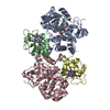
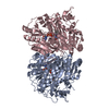
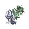
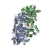
| Structure viewer | Molecule: MolmilJmol/JSmol |
|---|
- Downloads & links
-Download
| PDBx/mmCIF format | 1tup.cif.gz | 161.4 KB | Display | PDBx/mmCIF format |
|---|---|---|---|---|
| PDB format | pdb1tup.ent.gz | 122.3 KB | Display | PDB format |
| PDBx/mmJSON format | 1tup.json.gz | Tree view | PDBx/mmJSON format | |
| Others |  Other downloads Other downloads |
-Validation report
| Arichive directory | https://data.pdbj.org/pub/pdb/validation_reports/tu/1tupftp://data.pdbj.org/pub/pdb/validation_reports/tu/1tup | HTTPS FTP |
|---|
-Related structure data


-Links
 PDBj
PDBj


- Assembly
| Deposited unit | 
| ||||||||||||||||
|---|---|---|---|---|---|---|---|---|---|---|---|---|---|---|---|---|---|
| 1 |
| ||||||||||||||||
| Unit cell |
| ||||||||||||||||
| Noncrystallographic symmetry (NCS) | NCS oper:
|
-Components
| #1: DNA chain | Mass: 6348.121 Da / Num. of mol.: 1 / Source method: obtained synthetically | ||||
|---|---|---|---|---|---|
| #2: DNA chain | Mass: 6535.260 Da / Num. of mol.: 1 / Source method: obtained synthetically | ||||
| #3: Protein | Mass: 24586.877 Da / Num. of mol.: 3 Source method: isolated from a genetically manipulated source Source: (gene. exp.) Homo sapiens (human) / Cell: HUMAN VULVA CARCINOMA / Cell line: A431 / Plasmid: PET3D / Production host:  #4: Chemical |   Mass: 65.409 Da / Num. of mol.: 3 / Source method: obtained synthetically / Formula: Zn Mass: 65.409 Da / Num. of mol.: 3 / Source method: obtained synthetically / Formula: Zn#5: Water | ChemComp-HOH / |  Mass: 18.015 Da / Num. of mol.: 384 / Source method: isolated from a natural source / Formula: H2O Mass: 18.015 Da / Num. of mol.: 384 / Source method: isolated from a natural source / Formula: H2O |
-Experimental details
-Experiment
| Experiment | Method: X-RAY DIFFRACTION |
|---|
- Sample preparation
| Crystal | Density Matthews: 2.64 Å3/Da / Density % sol: 53.4 % | ||||||||||||||||||||||||||||||||||||||||||||||||||||||
|---|---|---|---|---|---|---|---|---|---|---|---|---|---|---|---|---|---|---|---|---|---|---|---|---|---|---|---|---|---|---|---|---|---|---|---|---|---|---|---|---|---|---|---|---|---|---|---|---|---|---|---|---|---|---|---|
| Crystal | *PLUS | ||||||||||||||||||||||||||||||||||||||||||||||||||||||
| Crystal grow | *PLUS Temperature: 4 ℃ / pH: 6.8 / Method: vapor diffusion, hanging drop | ||||||||||||||||||||||||||||||||||||||||||||||||||||||
| Components of the solutions | *PLUS
|
-Data collection
| Diffraction source | Wavelength: 1.54 |
|---|---|
| Detector | Type: RIGAKU RAXIS II / Detector: IMAGE PLATE |
| Radiation | Protocol: SINGLE WAVELENGTH / Monochromatic (M) / Laue (L): M / Scattering type: x-ray |
| Radiation wavelength | Wavelength: 1.54 Å / Relative weight: 1 |
| Reflection | Redundancy: 3.36 % / Rmerge(I) obs: 0.057 |
| Reflection | *PLUS Highest resolution: 2.2 Å / Num. obs: 36754 / % possible obs: 86 % / Observed criterion σ(I): 2 / Num. measured all: 123663 |
- Processing
| Software |
| ||||||||||||||||||||||||||||||||||||||||||||||||||||||||||||
|---|---|---|---|---|---|---|---|---|---|---|---|---|---|---|---|---|---|---|---|---|---|---|---|---|---|---|---|---|---|---|---|---|---|---|---|---|---|---|---|---|---|---|---|---|---|---|---|---|---|---|---|---|---|---|---|---|---|---|---|---|---|
| Refinement | Resolution: 2.2→6 Å / σ(F): 2 /
| ||||||||||||||||||||||||||||||||||||||||||||||||||||||||||||
| Refinement step | Cycle: LAST / Resolution: 2.2→6 Å
| ||||||||||||||||||||||||||||||||||||||||||||||||||||||||||||
| Refine LS restraints |
| ||||||||||||||||||||||||||||||||||||||||||||||||||||||||||||
| Refinement | *PLUS Highest resolution: 2.2 Å / Lowest resolution: 6 Å / σ(F): 2 | ||||||||||||||||||||||||||||||||||||||||||||||||||||||||||||
| Solvent computation | *PLUS | ||||||||||||||||||||||||||||||||||||||||||||||||||||||||||||
| Displacement parameters | *PLUS |
