 Movie
Movie Controller


+ Open data

- Basic information
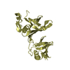
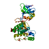
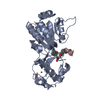
| Entry | Database: PDB / ID: 1ma3 | ||||||
|---|---|---|---|---|---|---|---|
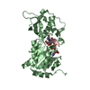
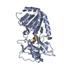
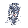
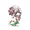
| Title | Structure of a Sir2 enzyme bound to an acetylated p53 peptide | ||||||
 Components Components |
| ||||||
 Keywords Keywords | PROTEIN BINDING / TRANSCRIPTION / ENZYME-SUBSTRATE COMPLEX | ||||||
| Function / homology |  Function and homology information Function and homology informationprotein-malonyllysine demalonylase activity / protein-succinyllysine desuccinylase activity / protein acetyllysine N-acetyltransferase / histone deacetylase activity, NAD-dependent / negative regulation of helicase activity / Loss of function of TP53 in cancer due to loss of tetramerization ability / Regulation of TP53 Expression / signal transduction by p53 class mediator / negative regulation of G1 to G0 transition / negative regulation of glucose catabolic process to lactate via pyruvate ...protein-malonyllysine demalonylase activity / protein-succinyllysine desuccinylase activity / protein acetyllysine N-acetyltransferase / histone deacetylase activity, NAD-dependent / negative regulation of helicase activity / Loss of function of TP53 in cancer due to loss of tetramerization ability / Regulation of TP53 Expression / signal transduction by p53 class mediator / negative regulation of G1 to G0 transition / negative regulation of glucose catabolic process to lactate via pyruvate / Transcriptional activation of cell cycle inhibitor p21 / regulation of intrinsic apoptotic signaling pathway by p53 class mediator / negative regulation of pentose-phosphate shunt / Activation of NOXA and translocation to mitochondria / ATP-dependent DNA/DNA annealing activity / regulation of cell cycle G2/M phase transition / oligodendrocyte apoptotic process / negative regulation of miRNA processing / intrinsic apoptotic signaling pathway in response to hypoxia / oxidative stress-induced premature senescence / regulation of tissue remodeling / positive regulation of thymocyte apoptotic process / positive regulation of mitochondrial membrane permeability / germ cell nucleus / regulation of fibroblast apoptotic process / bone marrow development / cellular response to actinomycin D / circadian behavior / histone deacetylase regulator activity / positive regulation of programmed necrotic cell death / : / regulation of mitochondrial membrane permeability involved in apoptotic process / RUNX3 regulates CDKN1A transcription / T cell proliferation involved in immune response / TP53 Regulates Transcription of Death Receptors and Ligands / Activation of PUMA and translocation to mitochondria / TP53 regulates transcription of additional cell cycle genes whose exact role in the p53 pathway remain uncertain / mRNA transcription / negative regulation of glial cell proliferation / negative regulation of neuroblast proliferation / regulation of DNA damage response, signal transduction by p53 class mediator / Regulation of TP53 Activity through Association with Co-factors / Formation of Senescence-Associated Heterochromatin Foci (SAHF) / mitochondrial DNA repair / T cell lineage commitment / thymocyte apoptotic process / ER overload response / TP53 Regulates Transcription of Caspase Activators and Caspases / cardiac septum morphogenesis / B cell lineage commitment / entrainment of circadian clock by photoperiod / negative regulation of DNA replication / Zygotic genome activation (ZGA) / negative regulation of mitophagy / TP53 Regulates Transcription of Genes Involved in Cytochrome C Release / necroptotic process / PI5P Regulates TP53 Acetylation / Association of TriC/CCT with target proteins during biosynthesis / positive regulation of release of cytochrome c from mitochondria / negative regulation of telomere maintenance via telomerase / SUMOylation of transcription factors / TP53 regulates transcription of several additional cell death genes whose specific roles in p53-dependent apoptosis remain uncertain / rRNA transcription / negative regulation of reactive oxygen species metabolic process / intrinsic apoptotic signaling pathway by p53 class mediator / TFIID-class transcription factor complex binding / Transcriptional Regulation by VENTX / cellular response to UV-C / viral process / neuroblast proliferation / NAD+ binding / intrinsic apoptotic signaling pathway in response to endoplasmic reticulum stress / replicative senescence / intrinsic apoptotic signaling pathway in response to DNA damage by p53 class mediator / Pyroptosis / positive regulation of RNA polymerase II transcription preinitiation complex assembly / chromosome organization / general transcription initiation factor binding / positive regulation of execution phase of apoptosis / hematopoietic stem cell differentiation / embryonic organ development / type II interferon-mediated signaling pathway / TP53 Regulates Transcription of Genes Involved in G1 Cell Cycle Arrest / response to X-ray / hematopoietic progenitor cell differentiation / somitogenesis / negative regulation of stem cell proliferation / core promoter sequence-specific DNA binding / glial cell proliferation / negative regulation of fibroblast proliferation / cis-regulatory region sequence-specific DNA binding / cellular response to glucose starvation / mitophagy / Regulation of TP53 Activity through Acetylation / negative regulation of proteolysis / mitotic G1 DNA damage checkpoint signaling / positive regulation of intrinsic apoptotic signaling pathway / response to salt stress / cardiac muscle cell apoptotic process / transcription repressor complex Similarity search - Function | ||||||
| Biological species |   Archaeoglobus fulgidus (archaea) Archaeoglobus fulgidus (archaea) | ||||||
| Method |  X-RAY DIFFRACTION / SYNCHROTRON / MOLECULAR REPLACEMENT / Resolution: 2 Å X-RAY DIFFRACTION / SYNCHROTRON / MOLECULAR REPLACEMENT / Resolution: 2 Å | ||||||
 Authors Authors | Avalos, J.L. / Celic, I. / Muhammad, S. / Cosgrove, M.S. / Boeke, J.D. / Wolberger, C. | ||||||
 Citation Citation | Journal: Mol.Cell / Year: 2002 Title: Structure of a Sir2 enzyme bound to an acetylated p53 peptide Authors: Avalos, J.L. / Celic, I. / Muhammad, S. / Cosgrove, M.S. / Boeke, J.D. / Wolberger, C. | ||||||
| History |
|
- Structure visualization
| Structure viewer | Molecule: MolmilJmol/JSmol |
|---|
- Downloads & links
-Download
| PDBx/mmCIF format | 1ma3.cif.gz | 67 KB | Display | PDBx/mmCIF format |
|---|---|---|---|---|
| PDB format | pdb1ma3.ent.gz | 47.9 KB | Display | PDB format |
| PDBx/mmJSON format | 1ma3.json.gz | Tree view | PDBx/mmJSON format | |
| Others |  Other downloads Other downloads |
-Validation report
| Arichive directory | https://data.pdbj.org/pub/pdb/validation_reports/ma/1ma3ftp://data.pdbj.org/pub/pdb/validation_reports/ma/1ma3 | HTTPS FTP |
|---|
-Related structure data
| Related structure data | 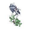 1iciS S: Starting model for refinement |
|---|---|
| Similar structure data |


-Links
 PDBj
PDBj


- Assembly
| Deposited unit | 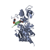
| ||||||||
|---|---|---|---|---|---|---|---|---|---|
| 1 |
| ||||||||
| Unit cell |
|
-Components
| #1: Protein | Mass: 28537.006 Da / Num. of mol.: 1 Source method: isolated from a genetically manipulated source Source: (gene. exp.) Archaeoglobus fulgidus (archaea) / Gene: SIR2-Af2 / Plasmid: pET11A / Production host:  |
|---|---|
| #2: Protein/peptide | Mass: 2139.521 Da / Num. of mol.: 1 / Fragment: Regulatory C-terminal tail (residues 372-389) / Source method: obtained synthetically Details: The sequence of the protein is naturally found in Homo sapiens. The protein is an Fmoc synthesized peptide. References: UniProt: P04637 |
| #3: Chemical | ChemComp-ZN /   Mass: 65.409 Da / Num. of mol.: 1 / Source method: obtained synthetically / Formula: Zn Mass: 65.409 Da / Num. of mol.: 1 / Source method: obtained synthetically / Formula: Zn |
| #4: Chemical | ChemComp-MES /   Mass: 195.237 Da / Num. of mol.: 1 / Source method: obtained synthetically / Formula: C6H13NO4S / Comment: pH buffer*YM Mass: 195.237 Da / Num. of mol.: 1 / Source method: obtained synthetically / Formula: C6H13NO4S / Comment: pH buffer*YM |
| #5: Water | ChemComp-HOH /  Mass: 18.015 Da / Num. of mol.: 129 / Source method: isolated from a natural source / Formula: H2O Mass: 18.015 Da / Num. of mol.: 129 / Source method: isolated from a natural source / Formula: H2O |
| Has protein modification | Y |
-Experimental details
-Experiment
| Experiment | Method: X-RAY DIFFRACTION / Number of used crystals: 1 |
|---|
- Sample preparation
| Crystal | Density Matthews: 1.98 Å3/Da / Density % sol: 37.79 % | |||||||||||||||||||||||||||||||||||||||||||||||||
|---|---|---|---|---|---|---|---|---|---|---|---|---|---|---|---|---|---|---|---|---|---|---|---|---|---|---|---|---|---|---|---|---|---|---|---|---|---|---|---|---|---|---|---|---|---|---|---|---|---|---|
| Crystal grow | Temperature: 291 K / Method: vapor diffusion, hanging drop / pH: 5.5 Details: PEG 8000, PEG 1000 and Sodium Chloride, pH 5.5, VAPOR DIFFUSION, HANGING DROP, temperature 291K | |||||||||||||||||||||||||||||||||||||||||||||||||
| Crystal grow | *PLUS Temperature: 20 ℃ | |||||||||||||||||||||||||||||||||||||||||||||||||
| Components of the solutions | *PLUS
|
-Data collection
| Diffraction | Mean temperature: 93 K |
|---|---|
| Diffraction source | Source: SYNCHROTRON / Site: NSLS  / Beamline: X25 / Wavelength: 1.1 Å / Beamline: X25 / Wavelength: 1.1 Å |
| Detector | Type: BRANDEIS - B4 / Detector: CCD / Date: Jul 20, 2001 |
| Radiation | Monochromator: Two crystal non-dispersive monochromator / Protocol: SINGLE WAVELENGTH / Monochromatic (M) / Laue (L): M / Scattering type: x-ray |
| Radiation wavelength | Wavelength: 1.1 Å / Relative weight: 1 |
| Reflection | Resolution: 2→50 Å / Num. all: 15490 / Num. obs: 15490 / % possible obs: 97.6 % / Observed criterion σ(I): -3 / Redundancy: 5.1 % / Biso Wilson estimate: 17.5 Å2 / Rmerge(I) obs: 0.101 / Rsym value: 0.101 / Net I/σ(I): 11.8 |
| Reflection shell | Highest resolution: 2 Å / Redundancy: 3.1 % / Rmerge(I) obs: 0.322 / Mean I/σ(I) obs: 4.2 / Num. unique all: 1414 / Rsym value: 0.323 / % possible all: 92.4 |
| Reflection | *PLUS Num. measured all: 79201 / Rmerge(I) obs: 0.113 |
| Reflection shell | *PLUS % possible obs: 92.4 % / Num. unique obs: 1414 / Num. measured obs: 4350 / Rmerge(I) obs: 0.342 |
- Processing
| Software |
| |||||||||||||||||||||||||
|---|---|---|---|---|---|---|---|---|---|---|---|---|---|---|---|---|---|---|---|---|---|---|---|---|---|---|
| Refinement | Method to determine structure: MOLECULAR REPLACEMENT Starting model: PDB entry 1ICI Resolution: 2→50 Å / Isotropic thermal model: isotropic / Cross valid method: THROUGHOUT / σ(F): 0 / Stereochemistry target values: Engh & Huber
| |||||||||||||||||||||||||
| Displacement parameters | Biso mean: 21.2 Å2 | |||||||||||||||||||||||||
| Refine analyze |
| |||||||||||||||||||||||||
| Refinement step | Cycle: LAST / Resolution: 2→50 Å
| |||||||||||||||||||||||||
| Refine LS restraints |
| |||||||||||||||||||||||||
| LS refinement shell | Highest resolution: 2 Å / Rfactor Rfree error: 0.05
| |||||||||||||||||||||||||
| Refinement | *PLUS % reflection Rfree: 5.17 % / Rfactor obs: 0.211 / Rfactor Rfree: 0.254 / Rfactor Rwork: 0.209 | |||||||||||||||||||||||||
| Solvent computation | *PLUS | |||||||||||||||||||||||||
| Displacement parameters | *PLUS | |||||||||||||||||||||||||
| Refine LS restraints | *PLUS
| |||||||||||||||||||||||||
| LS refinement shell | *PLUS Rfactor Rfree: 0.254 / Rfactor Rwork: 0.209 |
