 Movie
Movie Controller

[English] 日本語
 Yorodumi
- PDB-4utx: Crystal structure of zebrafish Sirtuin 5 in complex with 3-nitro-... -
+ Open data

- Basic information
| Entry | Database: PDB / ID: 4utx | ||||||
|---|---|---|---|---|---|---|---|
| Title | Crystal structure of zebrafish Sirtuin 5 in complex with 3-nitro- propionylated CPS1-peptide | ||||||
 Components Components |
| ||||||
 Keywords Keywords | HYDROLASE / REGULATORY ENZYME / DEACYLASE / MITOCHONDRIAL / ROSSMANN-FOLD / ZINC-BINDING | ||||||
| Function / homology |  Function and homology information Function and homology informationcarbamoyl phosphate biosynthetic process / cellular response to oleic acid / Transcriptional activation of mitochondrial biogenesis / monoatomic anion homeostasis / regulation of ketone biosynthetic process / peptidyl-lysine demalonylation / protein desuccinylation / peptidyl-lysine desuccinylation / protein-glutaryllysine deglutarylase activity / modified amino acid binding ...carbamoyl phosphate biosynthetic process / cellular response to oleic acid / Transcriptional activation of mitochondrial biogenesis / monoatomic anion homeostasis / regulation of ketone biosynthetic process / peptidyl-lysine demalonylation / protein desuccinylation / peptidyl-lysine desuccinylation / protein-glutaryllysine deglutarylase activity / modified amino acid binding / protein-malonyllysine demalonylase activity / protein-succinyllysine desuccinylase activity / carbamoyl-phosphate synthase (ammonia) / carbamoyl-phosphate synthase (ammonia) activity / carbamoyl-phosphate synthase (glutamine-hydrolyzing) activity / midgut development / triglyceride catabolic process / Urea cycle / homocysteine metabolic process / L-citrulline biosynthetic process / cellular response to ammonium ion / urea cycle / hepatocyte differentiation / histone deacetylase activity, NAD-dependent / L-glutamine metabolic process / response to growth hormone / response to amine / heterocyclic compound binding / response to alcohol / glutamate binding / response to zinc ion / response to starvation / response to dexamethasone / response to food / small molecule binding / potassium ion binding / acyl binding / mitochondrial nucleoid / NAD+ binding / response to amino acid / 'de novo' pyrimidine nucleobase biosynthetic process / nitric oxide metabolic process / cellular response to fibroblast growth factor stimulus / Transferases; Acyltransferases; Transferring groups other than aminoacyl groups / cellular response to glucagon stimulus / cellular response to cAMP / phospholipid binding / vasodilation / response to toxic substance / sperm principal piece / sperm midpiece / endopeptidase activity / response to lipopolysaccharide / mitochondrial inner membrane / mitochondrial matrix / response to xenobiotic stimulus / calcium ion binding / protein-containing complex binding / nucleolus / protein-containing complex / mitochondrion / zinc ion binding / ATP binding / metal ion binding / nucleus / plasma membrane / cytoplasm / cytosol Similarity search - Function | ||||||
| Biological species |   HOMO SAPIENS (human) HOMO SAPIENS (human) | ||||||
| Method |  X-RAY DIFFRACTION / SYNCHROTRON / MOLECULAR REPLACEMENT / Resolution: 3.1 Å X-RAY DIFFRACTION / SYNCHROTRON / MOLECULAR REPLACEMENT / Resolution: 3.1 Å | ||||||
 Authors Authors | Pannek, M. / Gertz, M. / Steegborn, C. | ||||||
 Citation Citation | Journal: Angew.Chem.Int.Ed.Engl. / Year: 2014 Title: Chemical Probing of the Human Sirtuin 5 Active Site Reveals its Substrate Acyl Specificity and Peptide-Based Inhibitors. Authors: Roessler, C. / Nowak, T. / Pannek, M. / Gertz, M. / Nguyen, G.T. / Scharfe, M. / Born, I. / Sippl, W. / Steegborn, C. / Schutkowski, M. | ||||||
| History |
|
- Structure visualization
| Structure viewer | Molecule: MolmilJmol/JSmol |
|---|
- Downloads & links
-Download
| PDBx/mmCIF format | 4utx.cif.gz | 216.3 KB | Display | PDBx/mmCIF format |
|---|---|---|---|---|
| PDB format | pdb4utx.ent.gz | 174.2 KB | Display | PDB format |
| PDBx/mmJSON format | 4utx.json.gz | Tree view | PDBx/mmJSON format | |
| Others |  Other downloads Other downloads |
-Validation report
| Arichive directory | https://data.pdbj.org/pub/pdb/validation_reports/ut/4utxftp://data.pdbj.org/pub/pdb/validation_reports/ut/4utx | HTTPS FTP |
|---|
-Related structure data
| Related structure data |  4utnC  4utrC  4utvC  4utzC  4uu7C  4uu8C  4uuaC  4uubC  2nyrS C: citing same article ( S: Starting model for refinement |
|---|---|
| Similar structure data |


-Links
 PDBj
PDBj


- Assembly
| Deposited unit | 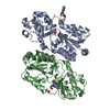
| ||||||||
|---|---|---|---|---|---|---|---|---|---|
| 1 | 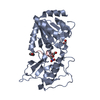
| ||||||||
| 2 | 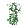
| ||||||||
| Unit cell |
| ||||||||
| Components on special symmetry positions |
| ||||||||
| Noncrystallographic symmetry (NCS) | NCS oper: (Code: given Matrix: (-0.75668, -0.5427, 0.36458), Vector: |
-Components
-Protein / Protein/peptide , 2 types, 3 molecules ABC
| #1: Protein | Mass: 30423.785 Da / Num. of mol.: 2 / Fragment: CATALYTIC CORE, UNP RESIDUES 30-298 Source method: isolated from a genetically manipulated source Source: (gene. exp.)  References: UniProt: Q6DHI5, Hydrolases; Acting on carbon-nitrogen bonds, other than peptide bonds; In linear amides #2: Protein/peptide | | Mass: 969.112 Da / Num. of mol.: 1 / Fragment: UNP RESIDUES 524-531 / Source method: obtained synthetically Details: BENZOYLATED GLYCINE AT POSITION 1. 3-NITRO-PROPIONYLATED LYSINE AT POSITION 4 Source: (synth.) HOMO SAPIENS (human) / References: UniProt: Q5R209, UniProt: P31327*PLUS |
|---|
-Non-polymers , 7 types, 43 molecules 


| #3: Chemical |  Mass: 65.409 Da / Num. of mol.: 2 / Source method: obtained synthetically / Formula: Zn Mass: 65.409 Da / Num. of mol.: 2 / Source method: obtained synthetically / Formula: Zn#4: Chemical |  Mass: 62.068 Da / Num. of mol.: 3 / Source method: obtained synthetically / Formula: C2H6O2 Mass: 62.068 Da / Num. of mol.: 3 / Source method: obtained synthetically / Formula: C2H6O2#5: Chemical | ChemComp-EPE / |  Mass: 238.305 Da / Num. of mol.: 1 / Source method: obtained synthetically / Formula: C8H18N2O4S / Comment: pH buffer*YM Mass: 238.305 Da / Num. of mol.: 1 / Source method: obtained synthetically / Formula: C8H18N2O4S / Comment: pH buffer*YM#6: Chemical | ChemComp-DMS / |  Mass: 78.133 Da / Num. of mol.: 1 / Source method: obtained synthetically / Formula: C2H6OS / Comment: DMSO, precipitant*YM Mass: 78.133 Da / Num. of mol.: 1 / Source method: obtained synthetically / Formula: C2H6OS / Comment: DMSO, precipitant*YM#7: Chemical | ChemComp-NA / |  Mass: 22.990 Da / Num. of mol.: 1 / Source method: obtained synthetically / Formula: Na Mass: 22.990 Da / Num. of mol.: 1 / Source method: obtained synthetically / Formula: Na#8: Chemical | ChemComp-3NP / |  Mass: 119.076 Da / Num. of mol.: 1 / Source method: obtained synthetically / Formula: C3H5NO4 Mass: 119.076 Da / Num. of mol.: 1 / Source method: obtained synthetically / Formula: C3H5NO4#9: Water | ChemComp-HOH / | Mass: 18.015 Da / Num. of mol.: 34 / Source method: isolated from a natural source / Formula: H2O |
|---|
-Details
| Sequence details | N-TERMINAL RESIDUES GIDPFT ARE ENCODED ON PET151 EXPRESSION |
|---|
-Experimental details
-Experiment
| Experiment | Method: X-RAY DIFFRACTION / Number of used crystals: 1 |
|---|
- Sample preparation
| Crystal | Density Matthews: 2.9 Å3/Da / Density % sol: 57 % / Description: NONE |
|---|---|
| Crystal grow | pH: 7.5 / Details: 22% PEG3350, 0.1 M HEPES PH 7.5 |
-Data collection
| Diffraction | Mean temperature: 100 K |
|---|---|
| Diffraction source | Source: SYNCHROTRON / Site: BESSY  / Beamline: 14.1 / Wavelength: 0.91705 / Beamline: 14.1 / Wavelength: 0.91705 |
| Detector | Type: DECTRIS PILATUS 6M / Detector: PIXEL / Date: Jul 5, 2013 / Details: MIRRORS |
| Radiation | Monochromator: SI-111 CRYSTAL / Protocol: SINGLE WAVELENGTH / Monochromatic (M) / Laue (L): M / Scattering type: x-ray |
| Radiation wavelength | Wavelength: 0.91705 Å / Relative weight: 1 |
| Reflection | Resolution: 3.1→50 Å / Num. obs: 13762 / % possible obs: 99.9 % / Observed criterion σ(I): 2 / Redundancy: 11.5 % / Rmerge(I) obs: 0.21 / Net I/σ(I): 14.3 |
| Reflection shell | Resolution: 3.1→3.2 Å / Redundancy: 10.6 % / Rmerge(I) obs: 1.31 / Mean I/σ(I) obs: 2.1 / % possible all: 100 |
- Processing
| Software |
| ||||||||||||||||||||||||||||||||||||||||||||||||||||||||||||||||||||||||||||||||||||||||||||||||||||||||||||||||||||||||||||||||||||||||||||||||||||||||||||||||||||||||||||||||||||||
|---|---|---|---|---|---|---|---|---|---|---|---|---|---|---|---|---|---|---|---|---|---|---|---|---|---|---|---|---|---|---|---|---|---|---|---|---|---|---|---|---|---|---|---|---|---|---|---|---|---|---|---|---|---|---|---|---|---|---|---|---|---|---|---|---|---|---|---|---|---|---|---|---|---|---|---|---|---|---|---|---|---|---|---|---|---|---|---|---|---|---|---|---|---|---|---|---|---|---|---|---|---|---|---|---|---|---|---|---|---|---|---|---|---|---|---|---|---|---|---|---|---|---|---|---|---|---|---|---|---|---|---|---|---|---|---|---|---|---|---|---|---|---|---|---|---|---|---|---|---|---|---|---|---|---|---|---|---|---|---|---|---|---|---|---|---|---|---|---|---|---|---|---|---|---|---|---|---|---|---|---|---|---|---|
| Refinement | Method to determine structure: MOLECULAR REPLACEMENT Starting model: PDB ENTRY 2NYR Resolution: 3.1→48.38 Å / Cor.coef. Fo:Fc: 0.941 / Cor.coef. Fo:Fc free: 0.886 / SU B: 45.245 / SU ML: 0.39 / Cross valid method: THROUGHOUT / ESU R Free: 0.469 / Stereochemistry target values: MAXIMUM LIKELIHOOD Details: HYDROGENS HAVE BEEN ADDED IN THE RIDING POSITIONS. U VALUES WITH TLS ADDED MISSING N-TERMINAL PROTEIN RESIDUES ARE DISORDERED. RESIDUES A280 TO A281 ARE DISORDERED. RESIDUES B275 TO B280 ARE ...Details: HYDROGENS HAVE BEEN ADDED IN THE RIDING POSITIONS. U VALUES WITH TLS ADDED MISSING N-TERMINAL PROTEIN RESIDUES ARE DISORDERED. RESIDUES A280 TO A281 ARE DISORDERED. RESIDUES B275 TO B280 ARE DISORDERED. PROTEIN RESIDUES CYS274 OF CHAIN A AND B FORM A DISULFIDE BRIDGE
| ||||||||||||||||||||||||||||||||||||||||||||||||||||||||||||||||||||||||||||||||||||||||||||||||||||||||||||||||||||||||||||||||||||||||||||||||||||||||||||||||||||||||||||||||||||||
| Solvent computation | Ion probe radii: 0.8 Å / Shrinkage radii: 0.8 Å / VDW probe radii: 1.2 Å / Solvent model: MASK | ||||||||||||||||||||||||||||||||||||||||||||||||||||||||||||||||||||||||||||||||||||||||||||||||||||||||||||||||||||||||||||||||||||||||||||||||||||||||||||||||||||||||||||||||||||||
| Displacement parameters | Biso mean: 67.991 Å2
| ||||||||||||||||||||||||||||||||||||||||||||||||||||||||||||||||||||||||||||||||||||||||||||||||||||||||||||||||||||||||||||||||||||||||||||||||||||||||||||||||||||||||||||||||||||||
| Refinement step | Cycle: LAST / Resolution: 3.1→48.38 Å
| ||||||||||||||||||||||||||||||||||||||||||||||||||||||||||||||||||||||||||||||||||||||||||||||||||||||||||||||||||||||||||||||||||||||||||||||||||||||||||||||||||||||||||||||||||||||
| Refine LS restraints |
|