 Movie
Movie Controller

[English] 日本語
 Yorodumi
- PDB-1olh: HIGH-RESOLUTION SOLUTION STRUCTURE OF THE OLIGOMERIZATION DOMAIN ... -
+ Open data

- Basic information
| Entry | Database: PDB / ID: 1olh | ||||||
|---|---|---|---|---|---|---|---|
| Title | HIGH-RESOLUTION SOLUTION STRUCTURE OF THE OLIGOMERIZATION DOMAIN OF P53 BY MULTI-DIMENSIONAL NMR | ||||||
 Components Components | TUMOR SUPPRESSOR P53 (OLIGOMERIZATION DOMAIN) | ||||||
 Keywords Keywords | ANTI-ONCOGENE PROTEIN | ||||||
| Function / homology |  Function and homology information Function and homology informationnegative regulation of helicase activity / Loss of function of TP53 in cancer due to loss of tetramerization ability / Regulation of TP53 Expression / signal transduction by p53 class mediator / negative regulation of G1 to G0 transition / Transcriptional activation of cell cycle inhibitor p21 / negative regulation of pentose-phosphate shunt / Activation of NOXA and translocation to mitochondria / ATP-dependent DNA/DNA annealing activity / regulation of cell cycle G2/M phase transition ...negative regulation of helicase activity / Loss of function of TP53 in cancer due to loss of tetramerization ability / Regulation of TP53 Expression / signal transduction by p53 class mediator / negative regulation of G1 to G0 transition / Transcriptional activation of cell cycle inhibitor p21 / negative regulation of pentose-phosphate shunt / Activation of NOXA and translocation to mitochondria / ATP-dependent DNA/DNA annealing activity / regulation of cell cycle G2/M phase transition / oligodendrocyte apoptotic process / positive regulation of thymocyte apoptotic process / oxidative stress-induced premature senescence / bone marrow development / circadian behavior / cellular response to actinomycin D / positive regulation of programmed necrotic cell death / RUNX3 regulates CDKN1A transcription / TP53 Regulates Transcription of Death Receptors and Ligands / Activation of PUMA and translocation to mitochondria / TP53 regulates transcription of additional cell cycle genes whose exact role in the p53 pathway remain uncertain / mRNA transcription / Urea cycle / Regulation of TP53 Activity through Association with Co-factors / ER overload response / hematopoietic stem cell differentiation / Formation of Senescence-Associated Heterochromatin Foci (SAHF) / TP53 Regulates Transcription of Caspase Activators and Caspases / intrinsic apoptotic signaling pathway by p53 class mediator / entrainment of circadian clock by photoperiod / Zygotic genome activation (ZGA) / TP53 Regulates Transcription of Genes Involved in Cytochrome C Release / PI5P Regulates TP53 Acetylation / positive regulation of release of cytochrome c from mitochondria / hematopoietic progenitor cell differentiation / Association of TriC/CCT with target proteins during biosynthesis / negative regulation of telomere maintenance via telomerase / SUMOylation of transcription factors / TP53 regulates transcription of several additional cell death genes whose specific roles in p53-dependent apoptosis remain uncertain / intrinsic apoptotic signaling pathway in response to DNA damage by p53 class mediator / Transcriptional Regulation by VENTX / replicative senescence / TFIID-class transcription factor complex binding / intrinsic apoptotic signaling pathway in response to endoplasmic reticulum stress / viral process / positive regulation of intrinsic apoptotic signaling pathway / Pyroptosis / determination of adult lifespan / positive regulation of RNA polymerase II transcription preinitiation complex assembly / negative regulation of fibroblast proliferation / general transcription initiation factor binding / positive regulation of execution phase of apoptosis / type II interferon-mediated signaling pathway / TP53 Regulates Transcription of Genes Involved in G1 Cell Cycle Arrest / cellular response to glucose starvation / core promoter sequence-specific DNA binding / cis-regulatory region sequence-specific DNA binding / Regulation of TP53 Activity through Acetylation / intrinsic apoptotic signaling pathway / mitotic G1 DNA damage checkpoint signaling / response to gamma radiation / 14-3-3 protein binding / MDM2/MDM4 family protein binding / TP53 Regulates Transcription of Genes Involved in G2 Cell Cycle Arrest / protein phosphatase 2A binding / molecular function activator activity / transcription initiation-coupled chromatin remodeling / Regulation of PTEN gene transcription / tumor necrosis factor-mediated signaling pathway / cellular response to ionizing radiation / cellular response to xenobiotic stimulus / DNA damage response, signal transduction by p53 class mediator / TP53 Regulates Metabolic Genes / autophagy / TP53 Regulates Transcription of DNA Repair Genes / Regulation of NF-kappa B signaling / mRNA 3'-UTR binding / protein tetramerization / promoter-specific chromatin binding / Stabilization of p53 / molecular condensate scaffold activity / negative regulation of cell growth / cellular response to gamma radiation / nucleotide-excision repair / G2/M Checkpoints / receptor tyrosine kinase binding / Autodegradation of the E3 ubiquitin ligase COP1 / PML body / positive regulation of miRNA transcription / DNA-binding transcription repressor activity, RNA polymerase II-specific / PKR-mediated signaling / Oncogene Induced Senescence / G2/M DNA damage checkpoint / Regulation of TP53 Activity through Methylation / cellular senescence / DNA Damage/Telomere Stress Induced Senescence / Pre-NOTCH Transcription and Translation / positive regulation of reactive oxygen species metabolic process / intracellular protein localization / histone deacetylase binding Similarity search - Function | ||||||
| Biological species |  Homo sapiens (human) Homo sapiens (human) | ||||||
| Method | SOLUTION NMR | ||||||
 Authors Authors | Clore, G.M. / Omichinski, J.G. / Gronenborn, A.M. | ||||||
 Citation Citation | Journal: Science / Year: 1994 Title: High-resolution structure of the oligomerization domain of p53 by multidimensional NMR. Authors: Clore, G.M. / Omichinski, J.G. / Sakaguchi, K. / Zambrano, N. / Sakamoto, H. / Appella, E. / Gronenborn, A.M. | ||||||
| History |
|
- Structure visualization
| Structure viewer | Molecule: MolmilJmol/JSmol |
|---|
- Downloads & links
-Download
| PDBx/mmCIF format | 1olh.cif.gz | 2 MB | Display | PDBx/mmCIF format |
|---|---|---|---|---|
| PDB format | pdb1olh.ent.gz | 1.7 MB | Display | PDB format |
| PDBx/mmJSON format | 1olh.json.gz | Tree view | PDBx/mmJSON format | |
| Others |  Other downloads Other downloads |
-Validation report
| Arichive directory | https://data.pdbj.org/pub/pdb/validation_reports/ol/1olhftp://data.pdbj.org/pub/pdb/validation_reports/ol/1olh | HTTPS FTP |
|---|
-Related structure data

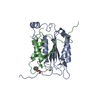
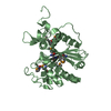
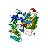
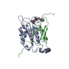
-Links
 PDBj
PDBj








- Assembly
| Deposited unit | 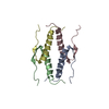
| |||||||||
|---|---|---|---|---|---|---|---|---|---|---|
| 1 |
| |||||||||
| NMR ensembles |
|
-Components
| #1: Protein/peptide | Mass: 4948.632 Da / Num. of mol.: 4 Source method: isolated from a genetically manipulated source Source: (gene. exp.) Homo sapiens (human) / References: UniProt: P04637 |
|---|
-Experimental details
-Experiment
| Experiment | Method: SOLUTION NMR |
|---|
- Processing
| Refinement | Software ordinal: 1 Details: THE 3D STRUCTURE OF THE OLIGOMERIZATION DOMAIN (RESIDUES 319 - 360) OF P53 BY MULTI-DIMENSIONAL HETERONUCLEAR-EDITED AND -FILTERED NMR IS BASED ON 3824 EXPERIMENTAL RESTRAINTS COMPRISING THE ...Details: THE 3D STRUCTURE OF THE OLIGOMERIZATION DOMAIN (RESIDUES 319 - 360) OF P53 BY MULTI-DIMENSIONAL HETERONUCLEAR-EDITED AND -FILTERED NMR IS BASED ON 3824 EXPERIMENTAL RESTRAINTS COMPRISING THE FOLLOWING INTRA- AND INTER-SUBUNIT RESTRAINTS: (A) INTRASUBUNIT: 840 SEQUENTIAL (|I-J|=1), 744 SHORT RANGE (1 < |I-J| >=5) AND 72 LONG RANGE (|I-J| >5) INTERRESIDUES AND INTRARESIDUE APPROXIMATE INTERPROTON DISTANCE RESTRAINTS, 136 DISTANCE RESTRAINTS FOR 68 HYDROGEN BONDS, 268 TORSION ANGLE (144 PHI, 104 CHI1 AND 20 CHI2) RESTRAINTS, AND 144 THREE-BOND HN-HA COUPLING CONSTANT RESTRAINTS. (B) INTERSUBUNIT: 96 A-B/C-D, 758 A-C/B-D, 10 A-D/B-C APPROXIMATE INTERPROTON DISTANCE RESTRAINTS, AND 24 DISTANCE RESTRAINTS FOR 12 HYDROGEN BONDS INVOLVING THE A-C/B-D SUBUNITS. IN ADDITION, THERE ARE A TOTAL OF 38 CALPHA AND 38 CB CHEMICAL SHIFT RESTRAINTS PER SUBUNIT THAT HAVE BEEN INCORPORATED INTO THE REFINEMENT [J. KUSZWESKI, J. QIN, A.M. GRONENBORN AND G.M. CLORE, J. MAGN RESON. SER IN PRESS (1994)] THE STRUCTURES ARE CALCULATED USING THE HYBRID METRIC MATRIX DISTANCE GEOMETRY-DYNAMICAL SIMULATED ANNEALING METHOD DESCRIBED BY: NILGES, M., CLORE, G.M. AND GRONENBORN, A.M. (1988) FEBS LETT. 29, 317-324. ALL STRUCTURAL STATISTICS ARE GIVEN IN THE JRNL REFERENCE. THE RESTRAINED MINIMIZED AVERAGE STRUCTURE (SA)R IS PRESENTED IN PROTEIN DATA BANK ENTRY 1OLG. THIS IS OBTAINED BY FIRST AVERAGING THE COORDINATES OF THE INDIVIDUAL 35 DYNAMICAL SIMULATED ANNEALING SA STRUCTURES BEST FITTED TO RESIDUES 324 - 356 OF ALL FOUR SUBUNITS, AND SUBJECTING THE RESULTING COORDINATES TO RESTRAINED MINIMIZATION. THE QUANTITY PRESENTED IN COLUMNS 61 - 66 IN THIS SET OF COORDINATES (THE B-FACTOR FIELD IN X-RAY STRUCTURES) GIVES THE AVERAGE RMS DIFFERENCE BETWEEN THE INDIVIDUAL SA STRUCTURES AND THE MEAN STRUCTURE. THE NUMBERS IN COLUMNS 61 - 66 OF THE INDIVIDUAL STRUCTURES HAVE NO MEANING. NOTE THAT RESIDUES 319 - 323 AT THE N-TERMINUS AND RESIDUES 357 - 360 AT THE C-TERMINUS ARE DISORDERED. |
|---|---|
| NMR ensemble | Conformers submitted total number: 35 |
