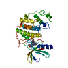
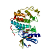
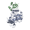
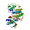
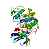
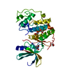
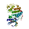
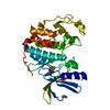
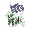
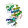
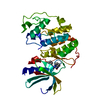
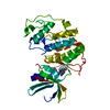
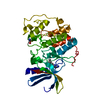
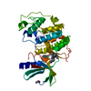
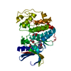
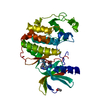
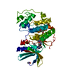
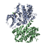
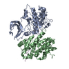
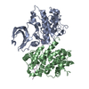
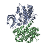
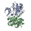
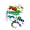
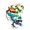
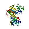
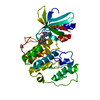
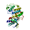
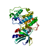
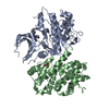
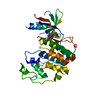
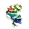
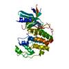
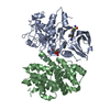
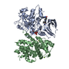
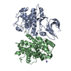
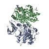
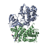
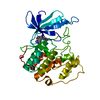
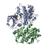
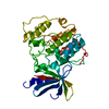
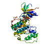
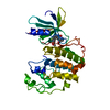
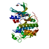
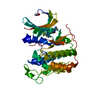
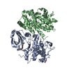
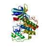
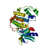
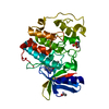
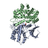
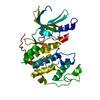
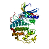
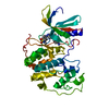
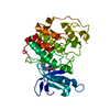
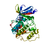
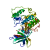
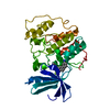
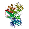
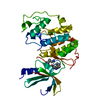
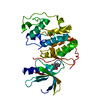
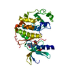
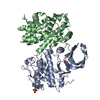
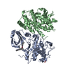
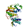
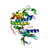
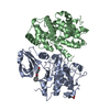
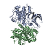
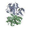
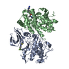
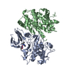
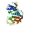
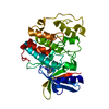
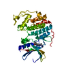
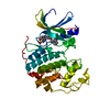
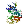
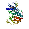
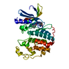
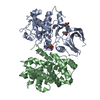
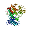
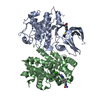
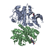
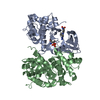
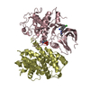
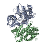
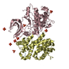
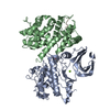
Entry Database : PDB / ID : 2jgzTitle Crystal structure of phospho-CDK2 in complex with Cyclin B CELL DIVISION PROTEIN KINASE 2 G2/MITOTIC-SPECIFIC CYCLIN-B1 Keywords / / / / / / / / / / / / / Function / homology Function Domain/homology Component
/ / / / / / / / / / / / / / / / / / / / / / / / / / / / / / / / / / / / / / / / / / / / / / / / / / / / / / / / / / / / / / / / / / / / / / / / / / / / / / / / / / / / / / / / / / / / / / / / / / / / / / / / / / / / / / / / / / / / / / / / / / / / / / / / / / / / / / / / / / / / / / / / / / / / / Biological species HOMO SAPIENS (human)Method / / / Resolution : 2.9 Å Authors Brown, N.R. / Petri, E. / Lowe, E.D. / Skamnaki, V. / Johnson, L.N. Journal : Cell Cycle / Year : 2007Title : Cyclin B and cyclin A confer different substrate recognition properties on CDK2.Authors : Brown, N.R. / Lowe, E.D. / Petri, E. / Skamnaki, V. / Antrobus, R. / Johnson, L.N. History Deposition Feb 17, 2007 Deposition site / Processing site Revision 1.0 May 22, 2007 Provider / Type Revision 1.1 May 8, 2011 Group Revision 1.2 Jul 13, 2011 Group Revision 1.3 Oct 9, 2019 Group Data collection / Database references ... Data collection / Database references / Derived calculations / Other Category / pdbx_database_status / struct_connItem _citation.journal_id_ISSN / _citation.page_last ... _citation.journal_id_ISSN / _citation.page_last / _citation.pdbx_database_id_DOI / _citation.title / _pdbx_database_status.status_code_sf / _struct_conn.pdbx_leaving_atom_flag Revision 1.4 Dec 13, 2023 Group / Database references / Refinement descriptionCategory chem_comp_atom / chem_comp_bond ... chem_comp_atom / chem_comp_bond / database_2 / pdbx_initial_refinement_model Item / _database_2.pdbx_database_accessionRevision 1.5 Oct 23, 2024 Group / Category / pdbx_modification_feature / Item
Show all Show less
 Movie
Movie Controller


 Open data
 Basic information
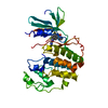
 Components
 Keywords
 Function and homology information
 HOMO SAPIENS (human)
 X-RAY DIFFRACTION /
  Authors
 Citation
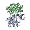
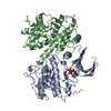
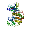
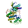
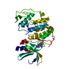
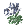
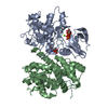
 Structure visualization
 Downloads & links
 Other downloads
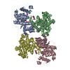


 PDBj
PDBj


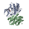
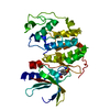
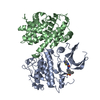
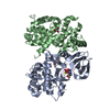
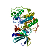
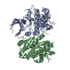
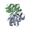
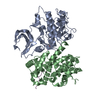
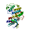
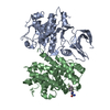
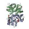
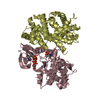
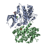
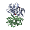
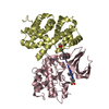
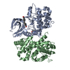
 Assembly
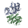

 Mass: 18.015 Da / Num. of mol.: 5 / Source method: isolated from a natural source / Formula: H2O
 Sample preparation
 / Beamline: ID29 / Wavelength: 0.8726
  Processing