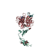
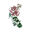
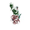
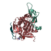
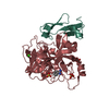
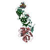
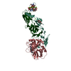
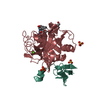
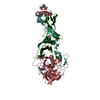
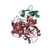
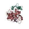
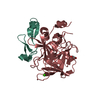
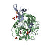
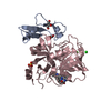
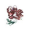
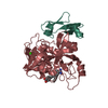
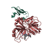
Entry Database : PDB / ID : 2bz6Title Orally available Factor7a inhibitor (BLOOD COAGULATION FACTOR ...) x 2 Keywords / / / Function / homology Function Domain/homology Component
/ / / / / / / / / / / / / / / / / / / / / / / / / / / / / / / / / / / / / / / / / / / / / / / / / / / / / / / / / / / / / / / / / / / / / / / / / / / / / / / / / / / / / / / / / / / / / / / / / Biological species HOMO SAPIENS (human)Method / / OTHER / Resolution : 1.6 Å Authors Groebke-Zbinden, K. / Obst-Sander, U. / Hilpert, K. / Kuehne, H. / Banner, D.W. / Boehm, H.J. / Stahl, M. / Ackermann, J. / Alig, L. / Weber, L. ...Groebke-Zbinden, K. / Obst-Sander, U. / Hilpert, K. / Kuehne, H. / Banner, D.W. / Boehm, H.J. / Stahl, M. / Ackermann, J. / Alig, L. / Weber, L. / Wessel, H.P. / Riederer, M.A. / Tschopp, T.B. / Lave, T. Journal : Bioorg.Med.Chem. / Year : 2006Title : Dose-Dependant Antithrombotic Activity of an Orally Active Tissue Factor/Factor Viia Inhibitor without Concomitant Enhancement of Bleeding Propensity.Authors : Groebke-Zbinden, K. / Banner, D.W. / Hilpert, K. / Himber, J. / Lave, T. / Riederer, M.A. / Stahl, M. / Tschopp, T.B. / Obst-Sander, U. History Deposition Aug 11, 2005 Deposition site / Processing site Revision 1.0 Feb 22, 2006 Provider / Type Revision 1.1 Jul 13, 2011 Group / Version format complianceRevision 1.2 Oct 9, 2024 Group Data collection / Database references ... Data collection / Database references / Derived calculations / Other / Structure summary Category chem_comp_atom / chem_comp_bond ... chem_comp_atom / chem_comp_bond / database_2 / pdbx_database_status / pdbx_entry_details / pdbx_modification_feature / pdbx_struct_conn_angle / struct_conn Item _database_2.pdbx_DOI / _database_2.pdbx_database_accession ... _database_2.pdbx_DOI / _database_2.pdbx_database_accession / _pdbx_database_status.status_code_sf / _pdbx_struct_conn_angle.ptnr1_auth_comp_id / _pdbx_struct_conn_angle.ptnr1_auth_seq_id / _pdbx_struct_conn_angle.ptnr1_label_asym_id / _pdbx_struct_conn_angle.ptnr1_label_atom_id / _pdbx_struct_conn_angle.ptnr1_label_comp_id / _pdbx_struct_conn_angle.ptnr1_label_seq_id / _pdbx_struct_conn_angle.ptnr3_auth_comp_id / _pdbx_struct_conn_angle.ptnr3_auth_seq_id / _pdbx_struct_conn_angle.ptnr3_label_asym_id / _pdbx_struct_conn_angle.ptnr3_label_atom_id / _pdbx_struct_conn_angle.ptnr3_label_comp_id / _pdbx_struct_conn_angle.ptnr3_label_seq_id / _pdbx_struct_conn_angle.value / _struct_conn.pdbx_dist_value / _struct_conn.ptnr1_auth_comp_id / _struct_conn.ptnr1_auth_seq_id / _struct_conn.ptnr1_label_asym_id / _struct_conn.ptnr1_label_atom_id / _struct_conn.ptnr1_label_comp_id / _struct_conn.ptnr1_label_seq_id / _struct_conn.ptnr2_auth_comp_id / _struct_conn.ptnr2_auth_seq_id / _struct_conn.ptnr2_label_asym_id / _struct_conn.ptnr2_label_atom_id / _struct_conn.ptnr2_label_comp_id / _struct_conn.ptnr2_label_seq_id
Show all Show less
 Movie
Movie Controller


 Open data
 Basic information
 Components
 Keywords
 Function and homology information
 HOMO SAPIENS (human)
 X-RAY DIFFRACTION /
  Authors
 Citation
 Structure visualization
 Downloads & links
 Other downloads


 PDBj
PDBj


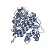
 Assembly
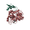

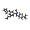


 Mass: 417.431 Da / Num. of mol.: 1 / Source method: obtained synthetically / Formula: C21H24FN3O5
 Mass: 96.063 Da / Num. of mol.: 1 / Source method: obtained synthetically / Formula: SO4
 Mass: 40.078 Da / Num. of mol.: 1 / Source method: obtained synthetically / Formula: Ca
 Sample preparation
 / Beamline: BM1A / Wavelength: 0.8
  Processing