Deposited unit
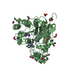
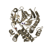
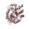
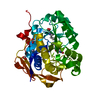
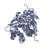
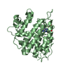
A: Protein phosphatase 2A, regulatory subunit B
B: Protein phosphatase 2A, regulatory subunit B
C: Protein phosphatase 2A, regulatory subunit B
D: Protein phosphatase 2A, regulatory subunit B
E: Protein phosphatase 2A, regulatory subunit B
F: Protein phosphatase 2A, regulatory subunit B
G: Protein phosphatase 2A, regulatory subunit B
H: Protein phosphatase 2A, regulatory subunit B
hetero molecules Summary Component details
Theoretical mass Number of molelcules Total (without water) 296,262 12 Polymers 294,553 8 Non-polymers 1,709 4 Water 0 0
1
A: Protein phosphatase 2A, regulatory subunit B Summary Component details Symmetry operations
Theoretical mass Number of molelcules Total (without water) 36,819 1 Polymers 36,819 1 Non-polymers 0 0 Water 0
Type Name Symmetry operation Number identity operation 1_555 x,y,z 1
2
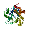
B: Protein phosphatase 2A, regulatory subunit B
hetero molecules Summary Component details Symmetry operations
Theoretical mass Number of molelcules Total (without water) 37,246 2 Polymers 36,819 1 Non-polymers 427 1 Water 0
Type Name Symmetry operation Number identity operation 1_555 x,y,z 1
3
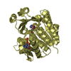
C: Protein phosphatase 2A, regulatory subunit B
hetero molecules Summary Component details Symmetry operations
Theoretical mass Number of molelcules Total (without water) 37,246 2 Polymers 36,819 1 Non-polymers 427 1 Water 0
Type Name Symmetry operation Number identity operation 1_555 x,y,z 1
4
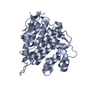
D: Protein phosphatase 2A, regulatory subunit B Summary Component details Symmetry operations
Theoretical mass Number of molelcules Total (without water) 36,819 1 Polymers 36,819 1 Non-polymers 0 0 Water 0
Type Name Symmetry operation Number identity operation 1_555 x,y,z 1
5
E: Protein phosphatase 2A, regulatory subunit B
hetero molecules Summary Component details Symmetry operations
Theoretical mass Number of molelcules Total (without water) 37,246 2 Polymers 36,819 1 Non-polymers 427 1 Water 0
Type Name Symmetry operation Number identity operation 1_555 x,y,z 1
6
F: Protein phosphatase 2A, regulatory subunit B Summary Component details Symmetry operations
Theoretical mass Number of molelcules Total (without water) 36,819 1 Polymers 36,819 1 Non-polymers 0 0 Water 0
Type Name Symmetry operation Number identity operation 1_555 x,y,z 1
7
G: Protein phosphatase 2A, regulatory subunit B
hetero molecules Summary Component details Symmetry operations
Theoretical mass Number of molelcules Total (without water) 37,246 2 Polymers 36,819 1 Non-polymers 427 1 Water 0
Type Name Symmetry operation Number identity operation 1_555 x,y,z 1
8
H: Protein phosphatase 2A, regulatory subunit B Summary Component details Symmetry operations
Theoretical mass Number of molelcules Total (without water) 36,819 1 Polymers 36,819 1 Non-polymers 0 0 Water 0
Type Name Symmetry operation Number identity operation 1_555 x,y,z 1
Unit cell Length a, b, c (Å) 80.041, 86.096, 95.099 Angle α, β, γ (deg.) 73.91, 89.97, 73.31 Int Tables number 1 Space group name H-M P1
Noncrystallographic symmetry (NCS) NCS domain ID Ens-ID Details (eV)1 1 A2 1 D3 1 F4 1 H1 2 B2 2 C3 2 E4 2 G1 3 B2 3 C3 3 E4 3 G
NCS domain segments Component-ID / Refine code
Dom-ID Ens-ID Beg auth comp-ID Beg label comp-ID End auth comp-ID End label comp-ID Auth asym-ID Label asym-ID Auth seq-ID Label seq-ID 1 1 ASNASNSERSERAA22 - 322 22 - 322 2 1 ASNASNSERSERDD22 - 322 22 - 322 3 1 ASNASNSERSERFF22 - 322 22 - 322 4 1 ASNASNSERSERHH22 - 322 22 - 322 1 2 ASNASNSERSERBB22 - 322 22 - 322 2 2 ASNASNSERSERCC22 - 322 22 - 322 3 2 ASNASNSERSEREE22 - 322 22 - 322 4 2 ASNASNSERSERGG22 - 322 22 - 322 1 3 ADPADPADPADPBI324 2 3 ADPADPADPADPCJ324 3 3 ADPADPADPADPEK324 4 3 ADPADPADPADPGL324
NCS ensembles
 Movie
Movie Controller

 Yorodumi
 Open data
 Basic information
 Components
 Keywords
 Function and homology information
 Homo sapiens (human)
 X-RAY DIFFRACTION /
  Authors
 Citation
 Structure visualization
 Downloads & links
 Other downloads


 PDBj
PDBj

 Assembly


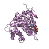


 Mass: 427.201 Da / Num. of mol.: 4 / Source method: obtained synthetically / Formula: C10H15N5O10P2 / Comment: ADP, energy-carrying molecule*YM
 Sample preparation
 / Beamline: X29A / Wavelength: 1.1 Å
 Processing