 Movie
Movie Controller

[English] 日本語
 Yorodumi
- PDB-1cvw: Crystal structure of active site-inhibited human coagulation fact... -
+ Open data

- Basic information
| Entry | Database: PDB / ID: 1cvw | ||||||
|---|---|---|---|---|---|---|---|
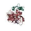
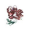
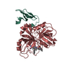
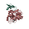
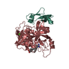
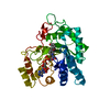
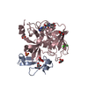
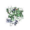
| Title | Crystal structure of active site-inhibited human coagulation factor VIIA (DES-GLA) | ||||||
 Components Components |
| ||||||
 Keywords Keywords | HYDROLASE/HYDROLASE INHIBITOR / BLOOD COAGULATION / FACTOR VIIA / SERINE PROTEASE / EGF / HYDROLASE-HYDROLASE INHIBITOR complex | ||||||
| Function / homology |  Function and homology information Function and homology informationcoagulation factor VIIa / response to Thyroid stimulating hormone / response to astaxanthin / response to thyrotropin-releasing hormone / response to 2,3,7,8-tetrachlorodibenzodioxine / response to carbon dioxide / serine-type peptidase complex / response to genistein / response to vitamin K / positive regulation of platelet-derived growth factor receptor signaling pathway ...coagulation factor VIIa / response to Thyroid stimulating hormone / response to astaxanthin / response to thyrotropin-releasing hormone / response to 2,3,7,8-tetrachlorodibenzodioxine / response to carbon dioxide / serine-type peptidase complex / response to genistein / response to vitamin K / positive regulation of platelet-derived growth factor receptor signaling pathway / positive regulation of leukocyte chemotaxis / response to thyroxine / response to cholesterol / positive regulation of positive chemotaxis / response to growth hormone / : / animal organ regeneration / positive regulation of blood coagulation / positive regulation of TOR signaling / Transport of gamma-carboxylated protein precursors from the endoplasmic reticulum to the Golgi apparatus / Gamma-carboxylation of protein precursors / Removal of aminoterminal propeptides from gamma-carboxylated proteins / BMAL1:CLOCK,NPAS2 activates circadian expression / serine-type peptidase activity / circadian rhythm / protein processing / response to estrogen / Golgi lumen / blood coagulation / response to estradiol / extracellular matrix / vesicle / response to hypoxia / positive regulation of cell migration / endoplasmic reticulum lumen / serine-type endopeptidase activity / signaling receptor binding / calcium ion binding / : / extracellular region / plasma membrane Similarity search - Function | ||||||
| Biological species |  Homo sapiens (human) Homo sapiens (human) | ||||||
| Method |  X-RAY DIFFRACTION / SYNCHROTRON / Resolution: 2.28 Å X-RAY DIFFRACTION / SYNCHROTRON / Resolution: 2.28 Å | ||||||
 Authors Authors | Kemball-Cook, G. / Johnson, D.J.D. / Tuddenham, E.G.D. / Harlos, K. | ||||||
 Citation Citation | Journal: J.Struct.Biol. / Year: 1999 Title: Crystal structure of active site-inhibited human coagulation factor VIIa (des-Gla). Authors: Kemball-Cook, G. / Johnson, D.J. / Tuddenham, E.G. / Harlos, K. #1: Journal: J.Struct.Biol. / Year: 1999Title: Crystallization and Preliminary X-ray Analysis of Active Site-Inhibited Human Coagulation Factor VIIa (des-Gla) Authors: Johnson, D.J. / Nugent, P.G. / Tuddenham, E.G. / Harlos, K. / Kemball-Cook, G. | ||||||
| History |
|
- Structure visualization
| Structure viewer | Molecule: MolmilJmol/JSmol |
|---|
- Downloads & links
-Download
| PDBx/mmCIF format | 1cvw.cif.gz | 80 KB | Display | PDBx/mmCIF format |
|---|---|---|---|---|
| PDB format | pdb1cvw.ent.gz | 58.2 KB | Display | PDB format |
| PDBx/mmJSON format | 1cvw.json.gz | Tree view | PDBx/mmJSON format | |
| Others |  Other downloads Other downloads |
-Validation report
| Arichive directory | https://data.pdbj.org/pub/pdb/validation_reports/cv/1cvwftp://data.pdbj.org/pub/pdb/validation_reports/cv/1cvw | HTTPS FTP |
|---|
-Related structure data
| Related structure data | |
|---|---|
| Similar structure data |


-Links
 PDBj
PDBj


- Assembly
| Deposited unit | 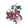
| ||||||||
|---|---|---|---|---|---|---|---|---|---|
| 1 |
| ||||||||
| Unit cell |
|
-Components
| #1: Protein | Mass: 6030.827 Da / Num. of mol.: 1 Source method: isolated from a genetically manipulated source Source: (gene. exp.) Homo sapiens (human) / Organ (production host): OVARY / Production host:   Cricetulus griseus (Chinese hamster) / References: UniProt: P08709, coagulation factor VIIa Cricetulus griseus (Chinese hamster) / References: UniProt: P08709, coagulation factor VIIa |
|---|---|
| #2: Protein | Mass: 28103.256 Da / Num. of mol.: 1 Source method: isolated from a genetically manipulated source Source: (gene. exp.) Homo sapiens (human) / Organ (production host): OVARY / Production host: Cricetulus griseus (Chinese hamster) / References: UniProt: P08709, coagulation factor VIIa |
| #3: Chemical | ChemComp-0GE /   Type: peptide-like, Peptide-like / Class: Inhibitor / Mass: 628.141 Da / Num. of mol.: 1 / Source method: obtained synthetically / Formula: C26H38ClN7O7S / References: dansyl-Glu-Gly-Arg chloromethyl ketone Type: peptide-like, Peptide-like / Class: Inhibitor / Mass: 628.141 Da / Num. of mol.: 1 / Source method: obtained synthetically / Formula: C26H38ClN7O7S / References: dansyl-Glu-Gly-Arg chloromethyl ketone |
| #4: Chemical | ChemComp-CA /   Mass: 40.078 Da / Num. of mol.: 1 / Source method: obtained synthetically / Formula: Ca Mass: 40.078 Da / Num. of mol.: 1 / Source method: obtained synthetically / Formula: Ca |
| #5: Water | ChemComp-HOH /  Mass: 18.015 Da / Num. of mol.: 171 / Source method: isolated from a natural source / Formula: H2O Mass: 18.015 Da / Num. of mol.: 171 / Source method: isolated from a natural source / Formula: H2O |
| Compound details | THE CHLOROMETHYLKETONE GROUP AND THE ADJACENT RESIDUE OF THE INHIBITOR BIND WITH PROTEIN BY TWO ...THE CHLOROMETH |
| Has protein modification | Y |
-Experimental details
-Experiment
| Experiment | Method: X-RAY DIFFRACTION / Number of used crystals: 1 |
|---|
- Sample preparation
| Crystal | Density Matthews: 3.72 Å3/Da / Density % sol: 66.98 % |
|---|---|
| Crystal grow | Temperature: 277 K / Method: vapor diffusion, sitting drop / pH: 7.5 Details: SOLUTION 30, HAMPTON SCREEN II, SEE REFERENCE 1, pH 7.5, VAPOR DIFFUSION, SITTING DROP, temperature 277K |
| Crystal grow | *PLUS Details: used micro bridge |
| Components of the solutions | *PLUS Conc.: 10 mg/ml / Common name: protein |
-Data collection
| Diffraction | Mean temperature: 100 K |
|---|---|
| Diffraction source | Source: SYNCHROTRON / Site: SRS  / Beamline: PX9.6 / Wavelength: 0.87 / Beamline: PX9.6 / Wavelength: 0.87 |
| Detector | Type: MARRESEARCH / Detector: IMAGE PLATE / Date: Jun 10, 1998 |
| Radiation | Protocol: SINGLE WAVELENGTH / Monochromatic (M) / Laue (L): M / Scattering type: x-ray |
| Radiation wavelength | Wavelength: 0.87 Å / Relative weight: 1 |
| Reflection | Resolution: 2.28→90 Å / Num. all: 23231 / Num. obs: 22596 / % possible obs: 92.3 % / Redundancy: 4.2 % / Rmerge(I) obs: 0.067 / Net I/σ(I): 22.8 |
| Reflection shell | Resolution: 2.28→2.36 Å / Redundancy: 2.2 % / Rmerge(I) obs: 0.294 / % possible all: 72.8 |
| Reflection | *PLUS Num. measured all: 345306 |
- Processing
| Software |
| ||||||||||||||||||||||||||||||||||||||||||||||||||||||||||||
|---|---|---|---|---|---|---|---|---|---|---|---|---|---|---|---|---|---|---|---|---|---|---|---|---|---|---|---|---|---|---|---|---|---|---|---|---|---|---|---|---|---|---|---|---|---|---|---|---|---|---|---|---|---|---|---|---|---|---|---|---|---|
| Refinement | Resolution: 2.28→30 Å / σ(F): 0 / Stereochemistry target values: ENGH & HUBER
| ||||||||||||||||||||||||||||||||||||||||||||||||||||||||||||
| Refinement step | Cycle: LAST / Resolution: 2.28→30 Å
| ||||||||||||||||||||||||||||||||||||||||||||||||||||||||||||
| Refine LS restraints |
| ||||||||||||||||||||||||||||||||||||||||||||||||||||||||||||
| Software | *PLUS Name: CNS / Classification: refinement | ||||||||||||||||||||||||||||||||||||||||||||||||||||||||||||
| Refinement | *PLUS Lowest resolution: 30 Å / σ(F): 0 | ||||||||||||||||||||||||||||||||||||||||||||||||||||||||||||
| Solvent computation | *PLUS | ||||||||||||||||||||||||||||||||||||||||||||||||||||||||||||
| Displacement parameters | *PLUS |
