Mass: 18.015 Da / Num. of mol.: 446 / Source method: isolated from a natural source / Formula: H2O
-
Details
Has protein modification
Y
-
Experimental details
-
Experiment
Experiment
Method: X-RAY DIFFRACTION / Number of used crystals: 1
-
Sample preparation
Crystal
Mosaicity: 0.783 °
Crystal grow
Temperature: 277 K / Method: vapor diffusion, sitting drop / pH: 8.5 Details: 16 mg/ml protein in 20mM Tris/HCl pH 8.4, 5 mM benzamidine, 0.1 M NaCl, 50 mM CaCl2 mixed 1+1 with 32-35% AMMONIUM SULPHATE, 2% PEG 4000, 0.1 M Bicine-NaOH pH 8.5, 15% glycerol
-
Data collection
Diffraction
Mean temperature: 100 K
Diffraction source
Source: ROTATING ANODE / Type: BRUKER AXS MICROSTAR / Wavelength: 1.54178 Å
Detector
Type: MAR scanner 345 mm plate / Detector: IMAGE PLATE / Date: Feb 19, 2008
Radiation
Protocol: SINGLE WAVELENGTH / Monochromatic (M) / Laue (L): M / Scattering type: x-ray
Radiation wavelength
Wavelength: 1.54178 Å / Relative weight: 1
Reflection
Resolution: 1.24→50 Å / Num. obs: 114725 / % possible obs: 75.9 % / Redundancy: 8 % / Biso Wilson estimate: 18.654 Å2 / Rmerge(I) obs: 0.056 / Χ2: 1.001 / Net I/av σ(I): 27.279 / Net I/σ(I): 13.2 / Num. measured all: 923374
Reflection shell
Diffraction-ID: 1 / Rejects: _
Resolution (Å)
Redundancy (%)
Num. unique all
% possible all
Χ2
Rmerge(I) obs
1.24-1.28
1
160
1.1
1.28-1.34
1.3
2458
16.5
1.02
1.34-1.4
2.2
7197
48.1
1.079
1.4-1.47
3.5
13509
90.1
0.972
0.877
1.47-1.56
6.6
15013
100
0.953
0.636
1.56-1.68
9.7
15024
100
0.992
0.354
1.68-1.85
9.9
15095
100
1.031
0.181
1.85-2.12
10.1
15178
100
1.01
0.094
2.12-2.67
10.2
15259
100
1.007
0.062
2.67-50
9.8
15832
99.9
0.997
0.038
-
Processing
Software
Name
Version
Classification
NB
SCALEPACK
datascaling
BUSTER
2.1.1
refinement
PDB_EXTRACT
3.22
dataextraction
XDS
datareduction
PHASER
phasing
Refinement
Method to determine structure: MOLECULAR REPLACEMENT Starting model: inhouse model Resolution: 1.4→33.65 Å / Cross valid method: THROUGHOUT / σ(F): 0 Details: the numbering follows that of the unprocessed precursor. the ligand may have occupancy less than 1. Trp424 flips underneath the 5-Hydroxy-1H-pyrazole. The main-chain around Trp424 has at ...Details: the numbering follows that of the unprocessed precursor. the ligand may have occupancy less than 1. Trp424 flips underneath the 5-Hydroxy-1H-pyrazole. The main-chain around Trp424 has at least two conformations and is largely disordered. several water molecules have been modeled with occupancies less than 1 and adopt mutually exclusive positions.
In the structure databanks used in Yorodumi, some data are registered as the other names, "COVID-19 virus" and "2019-nCoV". Here are the details of the virus and the list of structure data.
Jan 31, 2019. EMDB accession codes are about to change! (news from PDBe EMDB page)
EMDB accession codes are about to change! (news from PDBe EMDB page)
The allocation of 4 digits for EMDB accession codes will soon come to an end. Whilst these codes will remain in use, new EMDB accession codes will include an additional digit and will expand incrementally as the available range of codes is exhausted. The current 4-digit format prefixed with “EMD-” (i.e. EMD-XXXX) will advance to a 5-digit format (i.e. EMD-XXXXX), and so on. It is currently estimated that the 4-digit codes will be depleted around Spring 2019, at which point the 5-digit format will come into force.
The EM Navigator/Yorodumi systems omit the EMD- prefix.
Related info.:Q: What is EMD? / ID/Accession-code notation in Yorodumi/EM Navigator
Yorodumi is a browser for structure data from EMDB, PDB, SASBDB, etc.
This page is also the successor to EM Navigator detail page, and also detail information page/front-end page for Omokage search.
The word "yorodu" (or yorozu) is an old Japanese word meaning "ten thousand". "mi" (miru) is to see.
Related info.:EMDB / PDB / SASBDB / Comparison of 3 databanks / Yorodumi Search / Aug 31, 2016. New EM Navigator & Yorodumi / Yorodumi Papers / Jmol/JSmol / Function and homology information / Changes in new EM Navigator and Yorodumi
 Movie
Movie Controller

 Yorodumi
 Open data
 Basic information
 Components
 Keywords
 Function and homology information
 Homo sapiens (human)
 X-RAY DIFFRACTION /
  Authors
 Citation
 Structure visualization
 Downloads & links
 Other downloads


 PDBj
PDBj


 Assembly
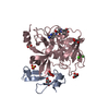


 Mass: 96.063 Da / Num. of mol.: 2 / Source method: obtained synthetically / Formula: SO4
 Mass: 92.094 Da / Num. of mol.: 5 / Source method: obtained synthetically / Formula: C3H8O3
 Mass: 40.078 Da / Num. of mol.: 1 / Source method: obtained synthetically / Formula: Ca
 Mass: 35.453 Da / Num. of mol.: 3 / Source method: obtained synthetically / Formula: Cl
 Mass: 295.296 Da / Num. of mol.: 1 / Source method: obtained synthetically / Formula: C15H13N5O2
 Sample preparation
 Processing