[English] 日本語
Yorodumi
- PDB-5pb2: Crystal Structure of Factor VIIa in complex with 2-phenyl-4-(1H-p... -

+
Open data


ID or keywords:

Loading...

-
Basic information

Entry
Database: PDB / ID: 5pb2
TitleCrystal Structure of Factor VIIa in complex with 2-phenyl-4-(1H-pyrrolo[3,2-c]pyridin-2-yl)pyrazol-3-ol
Components(Coagulation factor VII ...) x 2
KeywordsHYDROLASE/HYDROLASE INHIBITOR / GLYCOPROTEIN / HYDROLASE / SERINE PROTEASE / PLASMA / BLOOD COAGULATION FACTOR / PROTEIN INHIBITOR COMPLEX / CALCIUM-BINDING / HYDROLASE-HYDROLASE INHIBITOR COMPLEX
Function / homology
Function and homology information


coagulation factor VIIa / response to Thyroid stimulating hormone / response to 2,3,7,8-tetrachlorodibenzodioxine / response to astaxanthin / response to thyrotropin-releasing hormone / response to genistein / serine-type peptidase complex / positive regulation of platelet-derived growth factor receptor signaling pathway / response to vitamin K / response to carbon dioxide ...coagulation factor VIIa / response to Thyroid stimulating hormone / response to 2,3,7,8-tetrachlorodibenzodioxine / response to astaxanthin / response to thyrotropin-releasing hormone / response to genistein / serine-type peptidase complex / positive regulation of platelet-derived growth factor receptor signaling pathway / response to vitamin K / response to carbon dioxide / response to thyroxine / positive regulation of leukocyte chemotaxis / response to cholesterol / response to growth hormone / positive regulation of positive chemotaxis / Extrinsic Pathway of Fibrin Clot Formation / positive regulation of TOR signaling / animal organ regeneration / positive regulation of blood coagulation / Gamma-carboxylation of protein precursors / Transport of gamma-carboxylated protein precursors from the endoplasmic reticulum to the Golgi apparatus / Removal of aminoterminal propeptides from gamma-carboxylated proteins / serine-type peptidase activity / BMAL1:CLOCK,NPAS2 activates circadian gene expression / protein processing / Golgi lumen / circadian rhythm / response to estrogen / blood coagulation / response to estradiol / collagen-containing extracellular matrix / vesicle / response to hypoxia / positive regulation of cell migration / endoplasmic reticulum lumen / signaling receptor binding / serine-type endopeptidase activity / calcium ion binding / extracellular space / extracellular region / plasma membrane
Similarity search - Function
Peptidase S1A, coagulation factor VII/IX/X/C/Z / Coagulation factor-like, Gla domain superfamily / Coagulation Factor Xa inhibitory site / Laminin / Laminin / EGF-type aspartate/asparagine hydroxylation site / EGF-like domain / EGF-like calcium-binding, conserved site / Calcium-binding EGF-like domain signature. / Aspartic acid and asparagine hydroxylation site. ...Peptidase S1A, coagulation factor VII/IX/X/C/Z / Coagulation factor-like, Gla domain superfamily / Coagulation Factor Xa inhibitory site / Laminin / Laminin / EGF-type aspartate/asparagine hydroxylation site / EGF-like domain / EGF-like calcium-binding, conserved site / Calcium-binding EGF-like domain signature. / Aspartic acid and asparagine hydroxylation site. / EGF-like calcium-binding domain / Calcium-binding EGF-like domain / Vitamin K-dependent carboxylation/gamma-carboxyglutamic (GLA) domain / Gamma-carboxyglutamic acid-rich (GLA) domain / Gamma-carboxyglutamic acid-rich (GLA) domain superfamily / Vitamin K-dependent carboxylation domain. / Gla domain profile. / Domain containing Gla (gamma-carboxyglutamate) residues. / Epidermal growth factor-like domain. / EGF-like domain profile. / EGF-like domain signature 2. / EGF-like domain signature 1. / EGF-like domain / Ribbon / Serine proteases, trypsin family, histidine active site / Serine proteases, trypsin family, serine active site / Peptidase S1A, chymotrypsin family / Serine proteases, trypsin family, histidine active site. / Serine proteases, trypsin domain profile. / Trypsin-like serine protease / Serine proteases, trypsin family, serine active site. / Serine proteases, trypsin domain / Trypsin / Trypsin-like serine proteases / Thrombin, subunit H / Peptidase S1, PA clan, chymotrypsin-like fold / Peptidase S1, PA clan / Beta Barrel / Mainly Beta
Similarity search - Domain/homology
Chem-9RP / Coagulation factor VII
Similarity search - Component
Biological speciesHomo sapiens (human)
MethodX-RAY DIFFRACTION / SYNCHROTRON / MOLECULAR REPLACEMENT / Resolution: 1.45 Å
AuthorsStihle, M. / Mayweg, A. / Roever, S. / Rudolph, M.G.
CitationJournal: To be published
Title: Crystal Structure of a Factor VIIa complex
Authors: Mayweg, A. / Roever, S. / Rudolph, M.G.
History
DepositionNov 16, 2016Deposition site: RCSB / Processing site: RCSB
Revision 1.0Jun 21, 2017Provider: repository / Type: Initial release
Revision 1.1Feb 21, 2018Group: Structure summary / Category: pdbx_deposit_group / Item: _pdbx_deposit_group.group_type
Revision 1.2Nov 17, 2021Group: Database references / Derived calculations / Structure summary
Category: database_2 / pdbx_deposit_group / pdbx_struct_special_symmetry
Item: _database_2.pdbx_DOI / _database_2.pdbx_database_accession / _pdbx_deposit_group.group_description
Revision 1.3Apr 3, 2024Group: Data collection / Refinement description
Category: chem_comp_atom / chem_comp_bond / pdbx_initial_refinement_model

-
Structure visualization

Structure viewerMolecule:
MolmilJmol/JSmol

Downloads & links

-
Assembly

Deposited unit
A: Coagulation factor VII light chain
C: Coagulation factor VII heavy chain
hetero molecules


Theoretical massNumber of molelcules
Total (without water)35,7569
Polymers35,0882
Non-polymers6687
Water6,233346
1


  • Idetical with deposited unit
  • defined by author
TypeNameSymmetry operationNumber
identity operation1_555x,y,z1
Unit cell
Length a, b, c (Å)95.683, 95.683, 116.746
Angle α, β, γ (deg.)90.000, 90.000, 90.000
Int Tables number92
Space group name H-MP41212
Components on special symmetry positions
IDModelComponents
11C-860-

HOH

-
Components

-
Coagulation factor VII ... , 2 types, 2 molecules AC

#1: Protein Coagulation factor VII light chain / Proconvertin / Serum prothrombin conversion accelerator / SPCA


Mass: 6984.911 Da / Num. of mol.: 1
Source method: isolated from a genetically manipulated source
Source: (gene. exp.) Homo sapiens (human) / Gene: F7 / Production host: Escherichia coli (E. coli) / Strain (production host): BL21(DE3) / References: UniProt: P08709, coagulation factor VIIa
#2: Protein Coagulation factor VII heavy chain / Proconvertin / Serum prothrombin conversion accelerator / SPCA


Mass: 28103.256 Da / Num. of mol.: 1
Source method: isolated from a genetically manipulated source
Source: (gene. exp.) Homo sapiens (human) / Gene: F7 / Production host: Escherichia coli (E. coli) / Strain (production host): BL21(DE3) / References: UniProt: P08709, coagulation factor VIIa

-
Non-polymers , 6 types, 353 molecules

#3: Chemical ChemComp-GOL / GLYCEROL / GLYCERIN / PROPANE-1,2,3-TRIOL / Glycerol


Mass: 92.094 Da / Num. of mol.: 2 / Source method: obtained synthetically / Formula: C3H8O3
#4: Chemical ChemComp-CA / CALCIUM ION


Mass: 40.078 Da / Num. of mol.: 1 / Source method: obtained synthetically / Formula: Ca
#5: Chemical ChemComp-CL / CHLORIDE ION / Chloride


Mass: 35.453 Da / Num. of mol.: 2 / Source method: obtained synthetically / Formula: Cl
#6: Chemical ChemComp-SO4 / SULFATE ION / Sulfate


Mass: 96.063 Da / Num. of mol.: 1 / Source method: obtained synthetically / Formula: SO4
#7: Chemical ChemComp-9RP / 1-phenyl-4-(1H-pyrrolo[3,2-c]pyridin-2-yl)-1H-pyrazol-5-ol


Mass: 276.293 Da / Num. of mol.: 1 / Source method: obtained synthetically / Formula: C16H12N4O
#8: Water ChemComp-HOH / water / Water


Mass: 18.015 Da / Num. of mol.: 346 / Source method: isolated from a natural source / Formula: H2O

-
Experimental details

-
Experiment

ExperimentMethod: X-RAY DIFFRACTION / Number of used crystals: 1

-
Sample preparation

Crystal growTemperature: 277 K / Method: vapor diffusion, sitting drop / pH: 8.5
Details: 16 mg/ml protein in 20mM Tris/HCl pH 8.4, 5 mM benzamidine, 0.1 M NaCl, 50 mM CaCl2 mixed 1+1 with 32-35% AMMONIUM SULPHATE, 2% PEG 4000, 0.1 M Bicine-NaOH pH 8.5, 15% glycerol

-
Data collection

DiffractionMean temperature: 100 K
Diffraction sourceSource: SYNCHROTRON / Site: SLS / Beamline: X10SA / Wavelength: 0.99999 Å
DetectorType: MARMOSAIC 225 mm CCD / Detector: CCD / Date: Feb 25, 2008
RadiationProtocol: SINGLE WAVELENGTH / Monochromatic (M) / Laue (L): M / Scattering type: x-ray
Radiation wavelengthWavelength: 0.99999 Å / Relative weight: 1
ReflectionResolution: 1.45→34.51 Å / Num. obs: 94122 / % possible obs: 97.8 % / Redundancy: 6.68 % / Rmerge(I) obs: 0.075 / Rsym value: 0.075 / Net I/σ(I): 12.86
Reflection shellResolution: 1.45→1.55 Å / Redundancy: 4.19 % / Rmerge(I) obs: 0.506 / Mean I/σ(I) obs: 2.03 / Rsym value: 0.506 / Rejects: 0 / % possible all: 89.3

-
Processing

Software
NameVersionClassificationNB
REFMAC5.4.0067refinement
PDB_EXTRACT3.22data extraction
DENZOdata reduction
SADABSdata scaling
PHASERphasing
Refinement
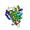
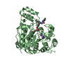
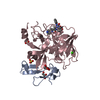
Method to determine structure: MOLECULAR REPLACEMENT
Starting model: inhouse model

Resolution: 1.45→33.83 Å / Cor.coef. Fo:Fc: 0.96 / Cor.coef. Fo:Fc free: 0.957 / SU B: 1.102 / SU ML: 0.042 / Cross valid method: THROUGHOUT / σ(F): 0 / ESU R: 0.059 / ESU R Free: 0.057 / Stereochemistry target values: MAXIMUM LIKELIHOOD
Details: The numbering follows that of the unprocessed precursor. There is density close (2A) to C8 of the azaindole moiety indicating that a small population of the S1-group could be rotated. The ...Details: The numbering follows that of the unprocessed precursor. There is density close (2A) to C8 of the azaindole moiety indicating that a small population of the S1-group could be rotated. The water molecule bridging the S1-group with Asp398 appears mobile. The binding mode induces strained geometry in Ser423, which is an outlier in the Ramachandran plot. The main-chain GLY425-GLY427 is largely disordered, GLN426 was modelled as ALA.. HYDROGENS HAVE BEEN ADDED IN THE RIDING POSITIONS
RfactorNum. reflection% reflectionSelection details
Rfree0.2036 4461 5 %RANDOM
Rwork0.1958 ---
obs0.1962 84967 93.02 %-
Solvent computationIon probe radii: 0.8 Å / Shrinkage radii: 0.8 Å / VDW probe radii: 1.2 Å / Solvent model: BABINET MODEL WITH MASK
Displacement parametersBiso max: 63.4 Å2 / Biso mean: 17.747 Å2 / Biso min: 10.56 Å2
Baniso -1Baniso -2Baniso -3
1--0.37 Å20 Å20 Å2
2---0.37 Å20 Å2
3---0.74 Å2
Refinement stepCycle: final / Resolution: 1.45→33.83 Å
ProteinNucleic acidLigandSolventTotal
Num. atoms2372 0 47 346 2765
Biso mean--31.84 33.97 -
Num. residues----308
Refine LS restraints
Refine-IDTypeDev idealDev ideal targetNumber
X-RAY DIFFRACTIONr_bond_refined_d0.0120.0212621
X-RAY DIFFRACTIONr_bond_other_d0.0020.021803
X-RAY DIFFRACTIONr_angle_refined_deg1.4681.973593
X-RAY DIFFRACTIONr_angle_other_deg2.6713.0074371
X-RAY DIFFRACTIONr_dihedral_angle_1_deg6.2025345
X-RAY DIFFRACTIONr_dihedral_angle_2_deg27.77922.655113
X-RAY DIFFRACTIONr_dihedral_angle_3_deg14.07415430
X-RAY DIFFRACTIONr_dihedral_angle_4_deg19.1761524
X-RAY DIFFRACTIONr_chiral_restr0.0810.2393
X-RAY DIFFRACTIONr_gen_planes_refined0.0060.0212946
X-RAY DIFFRACTIONr_gen_planes_other0.0020.02539
X-RAY DIFFRACTIONr_mcbond_it0.7761.51593
X-RAY DIFFRACTIONr_mcbond_other0.1331.5652
X-RAY DIFFRACTIONr_mcangle_it1.45522596
X-RAY DIFFRACTIONr_scbond_it1.79131028
X-RAY DIFFRACTIONr_scangle_it2.9984.5980
LS refinement shellResolution: 1.45→1.488 Å / Total num. of bins used: 20
RfactorNum. reflection% reflection
Rfree0.396 163 -
Rwork0.356 3482 -
all-3645 -
obs--52.19 %

+
About Yorodumi

-
News

-
Feb 9, 2022. New format data for meta-information of EMDB entries

New format data for meta-information of EMDB entries

  • Version 3 of the EMDB header file is now the official format.
  • The previous official version 1.9 will be removed from the archive.

Related info.:EMDB header

External links:wwPDB to switch to version 3 of the EMDB data model

-
Aug 12, 2020. Covid-19 info

Covid-19 info

URL: https://pdbj.org/emnavi/covid19.php

New page: Covid-19 featured information page in EM Navigator.

Related info.:Covid-19 info / Mar 5, 2020. Novel coronavirus structure data

+
Mar 5, 2020. Novel coronavirus structure data

Novel coronavirus structure data

Related info.:Yorodumi Speices / Aug 12, 2020. Covid-19 info

External links:COVID-19 featured content - PDBj / Molecule of the Month (242):Coronavirus Proteases

+
Jan 31, 2019. EMDB accession codes are about to change! (news from PDBe EMDB page)

EMDB accession codes are about to change! (news from PDBe EMDB page)

  • The allocation of 4 digits for EMDB accession codes will soon come to an end. Whilst these codes will remain in use, new EMDB accession codes will include an additional digit and will expand incrementally as the available range of codes is exhausted. The current 4-digit format prefixed with “EMD-” (i.e. EMD-XXXX) will advance to a 5-digit format (i.e. EMD-XXXXX), and so on. It is currently estimated that the 4-digit codes will be depleted around Spring 2019, at which point the 5-digit format will come into force.
  • The EM Navigator/Yorodumi systems omit the EMD- prefix.

Related info.:Q: What is EMD? / ID/Accession-code notation in Yorodumi/EM Navigator

External links:EMDB Accession Codes are Changing Soon! / Contact to PDBj

+
Jul 12, 2017. Major update of PDB

Major update of PDB

  • wwPDB released updated PDB data conforming to the new PDBx/mmCIF dictionary.
  • This is a major update changing the version number from 4 to 5, and with Remediation, in which all the entries are updated.
  • In this update, many items about electron microscopy experimental information are reorganized (e.g. em_software).
  • Now, EM Navigator and Yorodumi are based on the updated data.

External links:wwPDB Remediation / Enriched Model Files Conforming to OneDep Data Standards Now Available in the PDB FTP Archive

-
Yorodumi

Thousand views of thousand structures

  • Yorodumi is a browser for structure data from EMDB, PDB, SASBDB, etc.
  • This page is also the successor to EM Navigator detail page, and also detail information page/front-end page for Omokage search.
  • The word "yorodu" (or yorozu) is an old Japanese word meaning "ten thousand". "mi" (miru) is to see.

Related info.:EMDB / PDB / SASBDB / Comparison of 3 databanks / Yorodumi Search / Aug 31, 2016. New EM Navigator & Yorodumi / Yorodumi Papers / Jmol/JSmol / Function and homology information / Changes in new EM Navigator and Yorodumi

Read more