 Movie
Movie Controller


+ Open data

- Basic information
| Entry | Database: PDB / ID: 1kli | ||||||
|---|---|---|---|---|---|---|---|
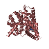
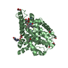
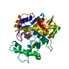
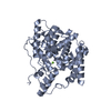
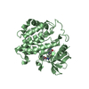
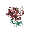
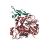
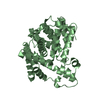
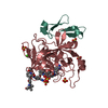
| Title | Cofactor-and substrate-assisted activation of factor VIIa | ||||||
 Components Components | (factor VIIa) x 2 | ||||||
 Keywords Keywords | HYDROLASE / extrinsic coagulation pathway / serine protease activation / rational drug design / substrate-assisted catalysis | ||||||
| Function / homology |  Function and homology information Function and homology informationcoagulation factor VIIa / response to Thyroid stimulating hormone / response to astaxanthin / response to thyrotropin-releasing hormone / response to 2,3,7,8-tetrachlorodibenzodioxine / response to carbon dioxide / serine-type peptidase complex / response to genistein / response to vitamin K / positive regulation of platelet-derived growth factor receptor signaling pathway ...coagulation factor VIIa / response to Thyroid stimulating hormone / response to astaxanthin / response to thyrotropin-releasing hormone / response to 2,3,7,8-tetrachlorodibenzodioxine / response to carbon dioxide / serine-type peptidase complex / response to genistein / response to vitamin K / positive regulation of platelet-derived growth factor receptor signaling pathway / positive regulation of leukocyte chemotaxis / response to thyroxine / response to cholesterol / positive regulation of positive chemotaxis / response to growth hormone / : / animal organ regeneration / positive regulation of blood coagulation / positive regulation of TOR signaling / Transport of gamma-carboxylated protein precursors from the endoplasmic reticulum to the Golgi apparatus / Gamma-carboxylation of protein precursors / Removal of aminoterminal propeptides from gamma-carboxylated proteins / BMAL1:CLOCK,NPAS2 activates circadian expression / serine-type peptidase activity / circadian rhythm / protein processing / response to estrogen / Golgi lumen / blood coagulation / response to estradiol / extracellular matrix / vesicle / response to hypoxia / positive regulation of cell migration / endoplasmic reticulum lumen / serine-type endopeptidase activity / signaling receptor binding / calcium ion binding / : / extracellular region / plasma membrane Similarity search - Function | ||||||
| Biological species |  Homo sapiens (human) Homo sapiens (human) | ||||||
| Method |  X-RAY DIFFRACTION / SYNCHROTRON / MOLECULAR REPLACEMENT / Resolution: 1.69 Å X-RAY DIFFRACTION / SYNCHROTRON / MOLECULAR REPLACEMENT / Resolution: 1.69 Å | ||||||
 Authors Authors | Sichler, K. / Banner, D.W. / D'Arcy, A. / Hopfner, K.P. / Huber, R. / Bode, W. / Kresse, G.B. / Kopetzki, E. / Brandstetter, H. | ||||||
 Citation Citation | Journal: J.Mol.Biol. / Year: 2002 Title: Crystal Structure of Uninhibited Factor VIIa Link its Cofactor and Substrate-assisted Activation to Specific Interactions Authors: Sichler, K. / Banner, D.W. / D'Arcy, A. / Hopfner, K.P. / Huber, R. / Bode, W. / Kresse, G.B. / Kopetzki, E. / Brandstetter, H. | ||||||
| History |
|
- Structure visualization
| Structure viewer | Molecule: MolmilJmol/JSmol |
|---|
- Downloads & links
-Download
| PDBx/mmCIF format | 1kli.cif.gz | 84.6 KB | Display | PDBx/mmCIF format |
|---|---|---|---|---|
| PDB format | pdb1kli.ent.gz | 62.2 KB | Display | PDB format |
| PDBx/mmJSON format | 1kli.json.gz | Tree view | PDBx/mmJSON format | |
| Others |  Other downloads Other downloads |
-Validation report
| Arichive directory | https://data.pdbj.org/pub/pdb/validation_reports/kl/1kliftp://data.pdbj.org/pub/pdb/validation_reports/kl/1kli | HTTPS FTP |
|---|
-Related structure data


-Links
 PDBj
PDBj


- Assembly
| Deposited unit | 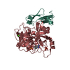
| ||||||||
|---|---|---|---|---|---|---|---|---|---|
| 1 |
| ||||||||
| 2 | 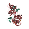
| ||||||||
| Unit cell |
|
-Components
-Protein , 2 types, 2 molecules LH
| #1: Protein | Mass: 7610.542 Da / Num. of mol.: 1 / Fragment: light chain Source method: isolated from a genetically manipulated source Details: N-terminal truncation / Source: (gene. exp.) Homo sapiens (human) / Species (production host): Escherichia coli / Production host:  |
|---|---|
| #2: Protein | Mass: 28103.256 Da / Num. of mol.: 1 / Fragment: heavy chain Source method: isolated from a genetically manipulated source Source: (gene. exp.) Homo sapiens (human) / Species (production host): Escherichia coli / Production host: |
-Non-polymers , 5 types, 306 molecules 


| #3: Chemical | ChemComp-SO4 /  Mass: 96.063 Da / Num. of mol.: 4 / Source method: obtained synthetically / Formula: SO4 Mass: 96.063 Da / Num. of mol.: 4 / Source method: obtained synthetically / Formula: SO4#4: Chemical | ChemComp-CA / |  Mass: 40.078 Da / Num. of mol.: 1 / Source method: obtained synthetically / Formula: Ca Mass: 40.078 Da / Num. of mol.: 1 / Source method: obtained synthetically / Formula: Ca#5: Chemical | ChemComp-BEN / |  Mass: 120.152 Da / Num. of mol.: 1 / Source method: obtained synthetically / Formula: C7H8N2 Mass: 120.152 Da / Num. of mol.: 1 / Source method: obtained synthetically / Formula: C7H8N2#6: Chemical | ChemComp-GOL / |  Mass: 92.094 Da / Num. of mol.: 1 / Source method: obtained synthetically / Formula: C3H8O3 Mass: 92.094 Da / Num. of mol.: 1 / Source method: obtained synthetically / Formula: C3H8O3#7: Water | ChemComp-HOH / | Mass: 18.015 Da / Num. of mol.: 299 / Source method: isolated from a natural source / Formula: H2O |
|---|
-Details
| Has protein modification | Y |
|---|
-Experimental details
-Experiment
| Experiment | Method: X-RAY DIFFRACTION / Number of used crystals: 1 |
|---|
- Sample preparation
| Crystal | Density Matthews: 3.57 Å3/Da / Density % sol: 65.52 % | ||||||||||||||||||||||||||||||||||||||||||
|---|---|---|---|---|---|---|---|---|---|---|---|---|---|---|---|---|---|---|---|---|---|---|---|---|---|---|---|---|---|---|---|---|---|---|---|---|---|---|---|---|---|---|---|
| Crystal grow | Temperature: 275 K / Method: vapor diffusion, sitting drop / pH: 8.5 Details: Ammonium sulphate, glycerol, PEG 400, BICINE, pH 8.5, VAPOR DIFFUSION, SITTING DROP, temperature 275K | ||||||||||||||||||||||||||||||||||||||||||
| Crystal grow | *PLUS Temperature: 4 ℃ | ||||||||||||||||||||||||||||||||||||||||||
| Components of the solutions | *PLUS
|
-Data collection
| Diffraction | Mean temperature: 170 K |
|---|---|
| Diffraction source | Source: SYNCHROTRON / Site: ESRF  / Beamline: BM1A / Wavelength: 1 Å / Beamline: BM1A / Wavelength: 1 Å |
| Detector | Type: ADSC QUANTUM 4 / Detector: CCD / Date: Jan 10, 2000 |
| Radiation | Protocol: SINGLE WAVELENGTH / Monochromatic (M) / Laue (L): M / Scattering type: x-ray |
| Radiation wavelength | Wavelength: 1 Å / Relative weight: 1 |
| Reflection | Resolution: 1.69→20 Å / Num. obs: 59659 / % possible obs: 98.3 % / Observed criterion σ(F): 2 / Observed criterion σ(I): 3 / Rmerge(I) obs: 0.057 |
| Reflection shell | Resolution: 1.69→1.75 Å / % possible all: 87.6 |
| Reflection | *PLUS % possible obs: 98.3 % / Redundancy: 9.33 % / Num. measured all: 556747 / Rmerge(I) obs: 0.057 |
| Reflection shell | *PLUS % possible obs: 87.6 % / Rmerge(I) obs: 0.652 |
- Processing
| Software |
| |||||||||||||||||||||||||
|---|---|---|---|---|---|---|---|---|---|---|---|---|---|---|---|---|---|---|---|---|---|---|---|---|---|---|
| Refinement | Method to determine structure: MOLECULAR REPLACEMENT / Resolution: 1.69→20 Å / σ(F): 0 / Stereochemistry target values: Engh & Huber
| |||||||||||||||||||||||||
| Refinement step | Cycle: LAST / Resolution: 1.69→20 Å
| |||||||||||||||||||||||||
| Refine LS restraints |
| |||||||||||||||||||||||||
| Refinement | *PLUS % reflection Rfree: 5 % / Rfactor Rfree: 0.2245 | |||||||||||||||||||||||||
| Solvent computation | *PLUS | |||||||||||||||||||||||||
| Displacement parameters | *PLUS | |||||||||||||||||||||||||
| Refine LS restraints | *PLUS
|
