 Movie
Movie Controller


+ Open data

- Basic information
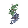
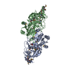
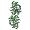
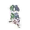
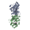
| Entry | Database: PDB / ID: 1j9c | ||||||
|---|---|---|---|---|---|---|---|
| Title | Crystal Structure of tissue factor-factor VIIa complex | ||||||
 Components Components |
| ||||||
 Keywords Keywords | HYDROLASE/HYDROLASE INHIBITOR / mobile loop / BLOOD CLOTTING / Hydrolase-hydrolase inhibitor complex | ||||||
| Function / homology |  Function and homology information Function and homology informationactivation of plasma proteins involved in acute inflammatory response / activation of blood coagulation via clotting cascade / coagulation factor VIIa / response to Thyroid stimulating hormone / response to astaxanthin / response to thyrotropin-releasing hormone / response to 2,3,7,8-tetrachlorodibenzodioxine / response to carbon dioxide / serine-type peptidase complex / response to genistein ...activation of plasma proteins involved in acute inflammatory response / activation of blood coagulation via clotting cascade / coagulation factor VIIa / response to Thyroid stimulating hormone / response to astaxanthin / response to thyrotropin-releasing hormone / response to 2,3,7,8-tetrachlorodibenzodioxine / response to carbon dioxide / serine-type peptidase complex / response to genistein / response to vitamin K / positive regulation of platelet-derived growth factor receptor signaling pathway / positive regulation of leukocyte chemotaxis / response to thyroxine / NGF-stimulated transcription / response to cholesterol / cytokine receptor activity / positive regulation of positive chemotaxis / : / response to growth hormone / positive regulation of blood coagulation / animal organ regeneration / positive regulation of endothelial cell apoptotic process / positive regulation of TOR signaling / Transport of gamma-carboxylated protein precursors from the endoplasmic reticulum to the Golgi apparatus / Gamma-carboxylation of protein precursors / Removal of aminoterminal propeptides from gamma-carboxylated proteins / positive regulation of endothelial cell proliferation / BMAL1:CLOCK,NPAS2 activates circadian expression / serine-type peptidase activity / positive regulation of interleukin-8 production / circadian rhythm / protein processing / phospholipid binding / response to estrogen / Golgi lumen / cytokine-mediated signaling pathway / positive regulation of angiogenesis / blood coagulation / response to estradiol / extracellular matrix / protease binding / vesicle / response to hypoxia / positive regulation of cell migration / endoplasmic reticulum lumen / signaling receptor binding / serine-type endopeptidase activity / external side of plasma membrane / calcium ion binding / positive regulation of gene expression / cell surface / : / extracellular region / membrane / plasma membrane Similarity search - Function | ||||||
| Biological species |  Homo sapiens (human) Homo sapiens (human) | ||||||
| Method |  X-RAY DIFFRACTION / SYNCHROTRON / MOLECULAR REPLACEMENT / Resolution: 2.9 Å X-RAY DIFFRACTION / SYNCHROTRON / MOLECULAR REPLACEMENT / Resolution: 2.9 Å | ||||||
 Authors Authors | Huang, M. / Ruf, W. / Edgington, T.S. / Wilson, I.A. | ||||||
 Citation Citation | Journal: To be Published Title: Ligand Induced Conformational Transitions of Tissue Factor. Crystal Structure of the Tissue Factor:Factor VIIa Complex. Authors: Huang, M. / Ruf, W. / Edgington, T.S. / Wilson, I.A. | ||||||
| History |
|
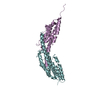
- Structure visualization
| Structure viewer | Molecule: MolmilJmol/JSmol |
|---|
- Downloads & links
-Download
| PDBx/mmCIF format | 1j9c.cif.gz | 121.4 KB | Display | PDBx/mmCIF format |
|---|---|---|---|---|
| PDB format | pdb1j9c.ent.gz | 92.1 KB | Display | PDB format |
| PDBx/mmJSON format | 1j9c.json.gz | Tree view | PDBx/mmJSON format | |
| Others |  Other downloads Other downloads |
-Validation report
| Arichive directory | https://data.pdbj.org/pub/pdb/validation_reports/j9/1j9cftp://data.pdbj.org/pub/pdb/validation_reports/j9/1j9c | HTTPS FTP |
|---|
-Related structure data
| Related structure data |  1danS S: Starting model for refinement |
|---|---|
| Similar structure data |


-Links
 PDBj
PDBj


- Assembly
| Deposited unit | 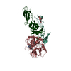
| ||||||||
|---|---|---|---|---|---|---|---|---|---|
| 1 |
| ||||||||
| Unit cell |
|
-Components
-Protein , 3 types, 3 molecules TLH
| #1: Protein | Mass: 23763.393 Da / Num. of mol.: 1 Source method: isolated from a genetically manipulated source Source: (gene. exp.) Homo sapiens (human) / Production host:  |
|---|---|
| #2: Protein | Mass: 10371.550 Da / Num. of mol.: 1 Source method: isolated from a genetically manipulated source Source: (gene. exp.) Homo sapiens (human) / Cell line (production host): BHK / Production host:   Cricetulus griseus (Chinese hamster) / References: UniProt: P08709 Cricetulus griseus (Chinese hamster) / References: UniProt: P08709 |
| #3: Protein | Mass: 28103.256 Da / Num. of mol.: 1 Source method: isolated from a genetically manipulated source Source: (gene. exp.) Homo sapiens (human) / Cell line (production host): BHK / Production host: Cricetulus griseus (Chinese hamster) / References: UniProt: P08709 |
-Sugars , 3 types, 3 molecules 

| #4: Sugar | ChemComp-GLC /  Type: D-saccharide, alpha linking / Mass: 180.156 Da / Num. of mol.: 1 Type: D-saccharide, alpha linking / Mass: 180.156 Da / Num. of mol.: 1Source method: isolated from a genetically manipulated source Formula: C6H12O6 |
|---|---|
| #5: Sugar | ChemComp-FUL /  Type: L-saccharide, beta linking / Mass: 164.156 Da / Num. of mol.: 1 Type: L-saccharide, beta linking / Mass: 164.156 Da / Num. of mol.: 1Source method: isolated from a genetically manipulated source Formula: C6H12O5 |
| #7: Sugar | ChemComp-NAG /  Type: D-saccharide, beta linking / Mass: 221.208 Da / Num. of mol.: 1 Type: D-saccharide, beta linking / Mass: 221.208 Da / Num. of mol.: 1Source method: isolated from a genetically manipulated source Formula: C8H15NO6 |
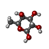
-Non-polymers , 3 types, 129 molecules 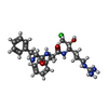


| #6: Chemical | ChemComp-0Z6 /  Type: peptide-like, Peptide-like / Class: Inhibitor / Mass: 504.045 Da / Num. of mol.: 1 / Source method: obtained synthetically / Formula: C25H36ClN6O3 / References: D-Phe-Phe-Arg Chloromethylketone Type: peptide-like, Peptide-like / Class: Inhibitor / Mass: 504.045 Da / Num. of mol.: 1 / Source method: obtained synthetically / Formula: C25H36ClN6O3 / References: D-Phe-Phe-Arg Chloromethylketone |
|---|---|
| #8: Chemical | ChemComp-CA /  Mass: 40.078 Da / Num. of mol.: 1 / Source method: obtained synthetically / Formula: Ca Mass: 40.078 Da / Num. of mol.: 1 / Source method: obtained synthetically / Formula: Ca |
| #9: Water | ChemComp-HOH / Mass: 18.015 Da / Num. of mol.: 127 / Source method: isolated from a natural source / Formula: H2O |
-Details
| Has protein modification | Y |
|---|---|
| Nonpolymer details | THE UNBOUND FORM OF THE INHIBITOR IS D-PHE-PHE-ARG-CHLOROMETHYLKETONE. UPON REACTION WITH PROTEIN ...THE UNBOUND FORM OF THE INHIBITOR IS D-PHE-PHE-ARG-CHLOROMETH |
-Experimental details
-Experiment
| Experiment | Method: X-RAY DIFFRACTION / Number of used crystals: 1 |
|---|
- Sample preparation
| Crystal | Density Matthews: 3.29 Å3/Da / Density % sol: 62.58 % |
|---|---|
| Crystal grow | Temperature: 295 K / Method: vapor diffusion, sitting drop / pH: 7.5 Details: 2% PEG 400, 2% 2-propanol, 2% PEG 4000, 12.5mM Calcium chloride, 2.0M ammonium sulfate, 0.1M HEPES, pH 7.5, VAPOR DIFFUSION, SITTING DROP, temperature 295K |
-Data collection
| Diffraction | Mean temperature: 177 K |
|---|---|
| Diffraction source | Source: SYNCHROTRON / Site: SSRL  / Beamline: BL7-1 / Wavelength: 1.08 Å / Beamline: BL7-1 / Wavelength: 1.08 Å |
| Detector | Type: MACSCIENCE / Detector: IMAGE PLATE / Date: Apr 4, 1999 Details: 58 cm long, Pt-coated, fused silica, vertical focusmirror |
| Radiation | Monochromator: Cyclindrically bent triangular Si(111) asymmetric cut, horizontal focus monochromator Protocol: SINGLE WAVELENGTH / Monochromatic (M) / Laue (L): M / Scattering type: x-ray |
| Radiation wavelength | Wavelength: 1.08 Å / Relative weight: 1 |
| Reflection | Resolution: 2.9→99 Å / Num. obs: 18285 / % possible obs: 99.9 % / Observed criterion σ(F): 0 / Observed criterion σ(I): 0 / Redundancy: 7.7 % / Biso Wilson estimate: -0.6 Å2 / Rmerge(I) obs: 0.119 / Net I/σ(I): 12.9 |
| Reflection shell | Resolution: 2.9→3 Å / Redundancy: 7.7 % / Rmerge(I) obs: 0.369 / % possible all: 100 |
- Processing
| Software |
| ||||||||||||||||||||||||
|---|---|---|---|---|---|---|---|---|---|---|---|---|---|---|---|---|---|---|---|---|---|---|---|---|---|
| Refinement | Method to determine structure: MOLECULAR REPLACEMENT Starting model: PDB ENTRY 1dan Resolution: 2.9→53.94 Å / Rfactor Rfree error: 0.013 / Data cutoff high absF: 275465.89 / Data cutoff low absF: 0 / Isotropic thermal model: RESTRAINED / Cross valid method: THROUGHOUT / σ(F): 0 / σ(I): 0 / Stereochemistry target values: Engh & Huber
| ||||||||||||||||||||||||
| Solvent computation | Solvent model: FLAT MODEL / Bsol: 47.46 Å2 / ksol: 0.344 e/Å3 | ||||||||||||||||||||||||
| Displacement parameters | Biso mean: 44.2 Å2
| ||||||||||||||||||||||||
| Refine analyze |
| ||||||||||||||||||||||||
| Refinement step | Cycle: LAST / Resolution: 2.9→53.94 Å
| ||||||||||||||||||||||||
| Refine LS restraints |
| ||||||||||||||||||||||||
| LS refinement shell | Resolution: 2.9→3.08 Å / Rfactor Rfree error: 0.043 / Total num. of bins used: 6
| ||||||||||||||||||||||||
| Xplor file |
|
