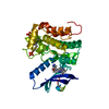
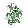
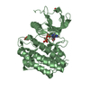
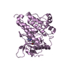
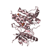
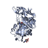
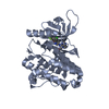
[English] 日本語
Yorodumi
- PDB-3pp0: Crystal Structure of the Kinase domain of Human HER2 (erbB2). -

+
Open data


ID or keywords:

Loading...

-
Basic information

Entry
Database: PDB / ID: 3pp0
TitleCrystal Structure of the Kinase domain of Human HER2 (erbB2).
ComponentsReceptor tyrosine-protein kinase erbB-2
KeywordsTRANSFERASE/TRANSFERASE INHIBITOR / KINASE DOMAIN / ANTI-ONCOGENE / ATP-BINDING / CELL CYCLE / DISEASE MUTATION / GLYCOPROTEIN / MEMBRANE / NUCLEOTIDE-BINDING / PHOSPHOPROTEIN / RECEPTOR / SECRETED / TRANSFERASE / TRANSMEMBRANE / TYROSINE-PROTEIN KINASE / TYROSINE KINASE INHIBITOR / TRANSFERASE-TRANSFERASE INHIBITOR complex
Function / homology
Function and homology information


negative regulation of immature T cell proliferation in thymus / ERBB3:ERBB2 complex / ERBB2-ERBB4 signaling pathway / GRB7 events in ERBB2 signaling / immature T cell proliferation in thymus / RNA polymerase I core binding / regulation of microtubule-based process / ErbB-3 class receptor binding / semaphorin receptor complex / Sema4D induced cell migration and growth-cone collapse ...negative regulation of immature T cell proliferation in thymus / ERBB3:ERBB2 complex / ERBB2-ERBB4 signaling pathway / GRB7 events in ERBB2 signaling / immature T cell proliferation in thymus / RNA polymerase I core binding / regulation of microtubule-based process / ErbB-3 class receptor binding / semaphorin receptor complex / Sema4D induced cell migration and growth-cone collapse / motor neuron axon guidance / neurotransmitter receptor localization to postsynaptic specialization membrane / PLCG1 events in ERBB2 signaling / neuromuscular junction development / ERBB2-EGFR signaling pathway / ERBB2 Activates PTK6 Signaling / positive regulation of Rho protein signal transduction / Drug-mediated inhibition of ERBB2 signaling / Resistance of ERBB2 KD mutants to trastuzumab / Resistance of ERBB2 KD mutants to sapitinib / Resistance of ERBB2 KD mutants to tesevatinib / Resistance of ERBB2 KD mutants to neratinib / Resistance of ERBB2 KD mutants to osimertinib / Resistance of ERBB2 KD mutants to afatinib / Resistance of ERBB2 KD mutants to AEE788 / Resistance of ERBB2 KD mutants to lapatinib / Drug resistance in ERBB2 TMD/JMD mutants / enzyme-linked receptor protein signaling pathway / positive regulation of transcription by RNA polymerase I / ERBB2-ERBB3 signaling pathway / oligodendrocyte differentiation / ERBB2 Regulates Cell Motility / semaphorin-plexin signaling pathway / PI3K events in ERBB2 signaling / positive regulation of cell adhesion / positive regulation of protein targeting to membrane / regulation of angiogenesis / coreceptor activity / Schwann cell development / Signaling by ERBB2 / cellular response to epidermal growth factor stimulus / cell surface receptor protein tyrosine kinase signaling pathway / GRB2 events in ERBB2 signaling / myelination / TFAP2 (AP-2) family regulates transcription of growth factors and their receptors / Downregulation of ERBB2:ERBB3 signaling / transmembrane receptor protein tyrosine kinase activity / SHC1 events in ERBB2 signaling / Constitutive Signaling by Overexpressed ERBB2 / phosphatidylinositol 3-kinase/protein kinase B signal transduction / neurogenesis / regulation of ERK1 and ERK2 cascade / basal plasma membrane / positive regulation of epithelial cell proliferation / positive regulation of translation / Signaling by ERBB2 TMD/JMD mutants / positive regulation of MAP kinase activity / wound healing / Signaling by ERBB2 ECD mutants / neuromuscular junction / neuron differentiation / Signaling by ERBB2 KD Mutants / receptor protein-tyrosine kinase / receptor tyrosine kinase binding / cellular response to growth factor stimulus / Downregulation of ERBB2 signaling / ruffle membrane / peptidyl-tyrosine phosphorylation / transmembrane signaling receptor activity / Constitutive Signaling by Aberrant PI3K in Cancer / PIP3 activates AKT signaling / myelin sheath / presynaptic membrane / heart development / PI5P, PP2A and IER3 Regulate PI3K/AKT Signaling / RAF/MAP kinase cascade / positive regulation of cell growth / basolateral plasma membrane / protein tyrosine kinase activity / positive regulation of MAPK cascade / cell surface receptor signaling pathway / early endosome / receptor complex / endosome membrane / intracellular signal transduction / positive regulation of protein phosphorylation / apical plasma membrane / protein heterodimerization activity / protein phosphorylation / signaling receptor binding / positive regulation of cell population proliferation / negative regulation of apoptotic process / perinuclear region of cytoplasm / signal transduction / nucleoplasm / ATP binding / identical protein binding / membrane / nucleus / plasma membrane
Similarity search - Function
: / Epidermal growth factor receptor transmembrane-juxtamembrane segment / Tyrosine protein kinase, EGF/ERB/XmrK receptor / Growth factor receptor domain 4 / Growth factor receptor domain IV / Receptor L-domain / Furin-like cysteine-rich domain / Receptor L-domain superfamily / Furin-like cysteine rich region / Receptor L domain ...: / Epidermal growth factor receptor transmembrane-juxtamembrane segment / Tyrosine protein kinase, EGF/ERB/XmrK receptor / Growth factor receptor domain 4 / Growth factor receptor domain IV / Receptor L-domain / Furin-like cysteine-rich domain / Receptor L-domain superfamily / Furin-like cysteine rich region / Receptor L domain / Furin-like repeat / Furin-like repeats / Growth factor receptor cysteine-rich domain superfamily / Tyrosine-protein kinase, catalytic domain / Tyrosine kinase, catalytic domain / Tyrosine protein kinases specific active-site signature. / Tyrosine-protein kinase, active site / Protein tyrosine and serine/threonine kinase / Serine-threonine/tyrosine-protein kinase, catalytic domain / Transferase(Phosphotransferase) domain 1 / Transferase(Phosphotransferase); domain 1 / Phosphorylase Kinase; domain 1 / Phosphorylase Kinase; domain 1 / Protein kinase, ATP binding site / Protein kinases ATP-binding region signature. / Protein kinase domain profile. / Protein kinase domain / Protein kinase-like domain superfamily / 2-Layer Sandwich / Orthogonal Bundle / Mainly Alpha / Alpha Beta
Similarity search - Domain/homology
Chem-03Q / Receptor tyrosine-protein kinase erbB-2
Similarity search - Component
Biological speciesHomo sapiens (human)
MethodX-RAY DIFFRACTION / SYNCHROTRON / MOLECULAR REPLACEMENT / Resolution: 2.25 Å
AuthorsSkene, R.J. / Aertgeerts, K. / Sogabe, S.
CitationJournal: J.Biol.Chem. / Year: 2011
Title: Structural Analysis of the Mechanism of Inhibition and Allosteric Activation of the Kinase Domain of HER2 Protein.
Authors: Aertgeerts, K. / Skene, R. / Yano, J. / Sang, B.C. / Zou, H. / Snell, G. / Jennings, A. / Iwamoto, K. / Habuka, N. / Hirokawa, A. / Ishikawa, T. / Tanaka, T. / Miki, H. / Ohta, Y. / Sogabe, S.
History
DepositionNov 23, 2010Deposition site: RCSB / Processing site: RCSB
Revision 1.0Mar 30, 2011Provider: repository / Type: Initial release
Revision 1.1Jul 13, 2011Group: Version format compliance
Revision 1.2Nov 8, 2017Group: Refinement description / Category: software / Item: _software.name
Revision 1.3Sep 6, 2023Group: Data collection / Database references ...Data collection / Database references / Derived calculations / Refinement description
Category: chem_comp_atom / chem_comp_bond ...chem_comp_atom / chem_comp_bond / database_2 / pdbx_initial_refinement_model / struct_ref_seq_dif / struct_site
Item: _database_2.pdbx_DOI / _database_2.pdbx_database_accession ..._database_2.pdbx_DOI / _database_2.pdbx_database_accession / _struct_ref_seq_dif.details / _struct_site.pdbx_auth_asym_id / _struct_site.pdbx_auth_comp_id / _struct_site.pdbx_auth_seq_id

-
Structure visualization

Structure viewerMolecule:
MolmilJmol/JSmol

Downloads & links

-
Assembly

Deposited unit
A: Receptor tyrosine-protein kinase erbB-2
B: Receptor tyrosine-protein kinase erbB-2
hetero molecules


Theoretical massNumber of molelcules
Total (without water)77,4464
Polymers76,4582
Non-polymers9882
Water3,423190
1
A: Receptor tyrosine-protein kinase erbB-2
hetero molecules


Theoretical massNumber of molelcules
Total (without water)38,7232
Polymers38,2291
Non-polymers4941
Water181
TypeNameSymmetry operationNumber
identity operation1_555x,y,z1
MethodPISA
2
B: Receptor tyrosine-protein kinase erbB-2
hetero molecules


Theoretical massNumber of molelcules
Total (without water)38,7232
Polymers38,2291
Non-polymers4941
Water181
TypeNameSymmetry operationNumber
identity operation1_555x,y,z1
MethodPISA
Unit cell
Length a, b, c (Å)48.705, 78.951, 152.675
Angle α, β, γ (deg.)90.00, 90.00, 90.00
Int Tables number19
Space group name H-MP212121

-
Components

#1: Protein Receptor tyrosine-protein kinase erbB-2 / Metastatic lymph node gene 19 protein / MLN 19 / Proto-oncogene Neu / Proto-oncogene c-ErbB-2 / ...Metastatic lymph node gene 19 protein / MLN 19 / Proto-oncogene Neu / Proto-oncogene c-ErbB-2 / Tyrosine kinase-type cell surface receptor HER2 / p185erbB2


Mass: 38229.012 Da / Num. of mol.: 2 / Fragment: kinase domain, residues 706-1009 / Mutation: M706A, Q711L, M712L
Source method: isolated from a genetically manipulated source
Source: (gene. exp.) Homo sapiens (human) / Gene: ERBB2, HER2, MLN19, NEU, NGL / Plasmid: pFastBac1 / Cell line (production host): Sf9 / Production host: 
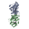
Spodoptera frugiperda (fall armyworm)
References: UniProt: P04626, receptor protein-tyrosine kinase
#2: Chemical ChemComp-03Q / 2-{2-[4-({5-chloro-6-[3-(trifluoromethyl)phenoxy]pyridin-3-yl}amino)-5H-pyrrolo[3,2-d]pyrimidin-5-yl]ethoxy}ethanol


Mass: 493.866 Da / Num. of mol.: 2 / Source method: obtained synthetically / Formula: C22H19ClF3N5O3
#3: Water ChemComp-HOH / water


Mass: 18.015 Da / Num. of mol.: 190 / Source method: isolated from a natural source / Formula: H2O

-
Experimental details

-
Experiment

ExperimentMethod: X-RAY DIFFRACTION / Number of used crystals: 1

-
Sample preparation

CrystalDensity Matthews: 1.92 Å3/Da / Density % sol: 35.92 %
Crystal growTemperature: 298 K / Method: vapor diffusion, sitting drop / pH: 6.5
Details: 20% PEG 3550, 200mM di-Ammonium Tartrate, 100 mM PIPES, pH 6.5, VAPOR DIFFUSION, SITTING DROP, temperature 298.0K

-
Data collection

DiffractionMean temperature: 100 K
Diffraction sourceSource: SYNCHROTRON / Site: ALS / Beamline: 5.0.3 / Wavelength: 1 Å
DetectorType: ADSC QUANTUM 315 / Detector: CCD / Date: Oct 28, 2005
RadiationMonochromator: Si (220) Single mono / Protocol: SINGLE WAVELENGTH / Monochromatic (M) / Laue (L): M / Scattering type: x-ray
Radiation wavelengthWavelength: 1 Å / Relative weight: 1
ReflectionResolution: 2.25→50 Å / Num. obs: 28773 / % possible obs: 99.5 % / Redundancy: 3.9 % / Rmerge(I) obs: 0.08 / Net I/σ(I): 15.1
Reflection shellResolution: 2.25→2.33 Å / Redundancy: 3.6 % / Rmerge(I) obs: 0.553 / Mean I/σ(I) obs: 1.82 / % possible all: 96.6

-
Processing

Software
NameVersionClassification
Blu-Icedata collection
MOLREPphasing
REFMAC5.5.0109refinement
HKL-2000data reduction
HKL-2000data scaling
RefinementMethod to determine structure: MOLECULAR REPLACEMENT
Starting model: 1M14
Resolution: 2.25→50 Å / Cor.coef. Fo:Fc: 0.957 / Cor.coef. Fo:Fc free: 0.92 / SU B: 17.529 / SU ML: 0.193 / Cross valid method: THROUGHOUT / ESU R Free: 0.255 / Stereochemistry target values: MAXIMUM LIKELIHOOD
RfactorNum. reflection% reflectionSelection details
Rfree0.26035 1438 5.1 %RANDOM
Rwork0.18484 ---
obs0.18865 27014 99.16 %-
Solvent computationIon probe radii: 0.8 Å / Shrinkage radii: 0.8 Å / VDW probe radii: 1.4 Å / Solvent model: MASK
Displacement parametersBiso mean: 42.366 Å2
Baniso -1Baniso -2Baniso -3
1-3.38 Å20 Å20 Å2
2---0.5 Å20 Å2
3----2.88 Å2
Refinement stepCycle: LAST / Resolution: 2.25→50 Å
ProteinNucleic acidLigandSolventTotal
Num. atoms4657 0 68 190 4915
Refine LS restraints
Refine-IDTypeDev idealDev ideal targetNumber
X-RAY DIFFRACTIONr_bond_refined_d0.010.0224837
X-RAY DIFFRACTIONr_bond_other_d
X-RAY DIFFRACTIONr_angle_refined_deg1.271.9926553
X-RAY DIFFRACTIONr_angle_other_deg
X-RAY DIFFRACTIONr_dihedral_angle_1_deg5.9495579
X-RAY DIFFRACTIONr_dihedral_angle_2_deg38.17822.981208
X-RAY DIFFRACTIONr_dihedral_angle_3_deg16.6115869
X-RAY DIFFRACTIONr_dihedral_angle_4_deg17.0911545
X-RAY DIFFRACTIONr_chiral_restr0.0870.2728
X-RAY DIFFRACTIONr_gen_planes_refined0.0050.0213598
X-RAY DIFFRACTIONr_gen_planes_other
X-RAY DIFFRACTIONr_nbd_refined
X-RAY DIFFRACTIONr_nbd_other
X-RAY DIFFRACTIONr_nbtor_refined
X-RAY DIFFRACTIONr_nbtor_other
X-RAY DIFFRACTIONr_xyhbond_nbd_refined
X-RAY DIFFRACTIONr_xyhbond_nbd_other
X-RAY DIFFRACTIONr_metal_ion_refined
X-RAY DIFFRACTIONr_metal_ion_other
X-RAY DIFFRACTIONr_symmetry_vdw_refined
X-RAY DIFFRACTIONr_symmetry_vdw_other
X-RAY DIFFRACTIONr_symmetry_hbond_refined
X-RAY DIFFRACTIONr_symmetry_hbond_other
X-RAY DIFFRACTIONr_symmetry_metal_ion_refined
X-RAY DIFFRACTIONr_symmetry_metal_ion_other
X-RAY DIFFRACTIONr_mcbond_it2.01222907
X-RAY DIFFRACTIONr_mcbond_other
X-RAY DIFFRACTIONr_mcangle_it3.3634726
X-RAY DIFFRACTIONr_scbond_it5.46741930
X-RAY DIFFRACTIONr_scangle_it7.86261826
X-RAY DIFFRACTIONr_rigid_bond_restr
X-RAY DIFFRACTIONr_sphericity_free
X-RAY DIFFRACTIONr_sphericity_bonded
LS refinement shellResolution: 2.252→2.31 Å / Total num. of bins used: 20
RfactorNum. reflection% reflection
Rfree0.371 104 -
Rwork0.266 1893 -
obs--95.78 %
Refinement TLS params.

Method: refined / Refine-ID: X-RAY DIFFRACTION

IDL112)L122)L132)L222)L232)L332)S11 (Å °)S12 (Å °)S13 (Å °)S21 (Å °)S22 (Å °)S23 (Å °)S31 (Å °)S32 (Å °)S33 (Å °)T112)T122)T132)T222)T232)T332)Origin x (Å)Origin y (Å)Origin z (Å)
10.8172-0.3151-0.75491.10470.20953.24720.0255-0.0362-0.0746-0.15360.03040.028-0.0897-0.0576-0.05590.04920-0.0220.0129-0.00170.044512.479921.62534.0645
21.02690.0926-0.31310.7641-0.39152.59670.04460.01780.0333-0.03180.0039-0.03540.08810.0031-0.04850.03880.00260.00690.0181-0.03120.062430.100840.4937-4.086
Refinement TLS group
IDRefine-IDRefine TLS-IDAuth asym-IDAuth seq-ID
1X-RAY DIFFRACTION1A706 - 1009
2X-RAY DIFFRACTION1A1
3X-RAY DIFFRACTION2B706 - 1009
4X-RAY DIFFRACTION2B2

+
About Yorodumi

-
News

-
Feb 9, 2022. New format data for meta-information of EMDB entries

New format data for meta-information of EMDB entries

  • Version 3 of the EMDB header file is now the official format.
  • The previous official version 1.9 will be removed from the archive.

Related info.:EMDB header

External links:wwPDB to switch to version 3 of the EMDB data model

-
Aug 12, 2020. Covid-19 info

Covid-19 info

URL: https://pdbjlvh1.pdbj.org/emnavi/covid19.php

New page: Covid-19 featured information page in EM Navigator.

Related info.:Covid-19 info / Mar 5, 2020. Novel coronavirus structure data

+
Mar 5, 2020. Novel coronavirus structure data

Novel coronavirus structure data

Related info.:Yorodumi Speices / Aug 12, 2020. Covid-19 info

External links:COVID-19 featured content - PDBj / Molecule of the Month (242):Coronavirus Proteases

+
Jan 31, 2019. EMDB accession codes are about to change! (news from PDBe EMDB page)

EMDB accession codes are about to change! (news from PDBe EMDB page)

  • The allocation of 4 digits for EMDB accession codes will soon come to an end. Whilst these codes will remain in use, new EMDB accession codes will include an additional digit and will expand incrementally as the available range of codes is exhausted. The current 4-digit format prefixed with “EMD-” (i.e. EMD-XXXX) will advance to a 5-digit format (i.e. EMD-XXXXX), and so on. It is currently estimated that the 4-digit codes will be depleted around Spring 2019, at which point the 5-digit format will come into force.
  • The EM Navigator/Yorodumi systems omit the EMD- prefix.

Related info.:Q: What is EMD? / ID/Accession-code notation in Yorodumi/EM Navigator

External links:EMDB Accession Codes are Changing Soon! / Contact to PDBj

+
Jul 12, 2017. Major update of PDB

Major update of PDB

  • wwPDB released updated PDB data conforming to the new PDBx/mmCIF dictionary.
  • This is a major update changing the version number from 4 to 5, and with Remediation, in which all the entries are updated.
  • In this update, many items about electron microscopy experimental information are reorganized (e.g. em_software).
  • Now, EM Navigator and Yorodumi are based on the updated data.

External links:wwPDB Remediation / Enriched Model Files Conforming to OneDep Data Standards Now Available in the PDB FTP Archive

-
Yorodumi

Thousand views of thousand structures

  • Yorodumi is a browser for structure data from EMDB, PDB, SASBDB, etc.
  • This page is also the successor to EM Navigator detail page, and also detail information page/front-end page for Omokage search.
  • The word "yorodu" (or yorozu) is an old Japanese word meaning "ten thousand". "mi" (miru) is to see.

Related info.:EMDB / PDB / SASBDB / Comparison of 3 databanks / Yorodumi Search / Aug 31, 2016. New EM Navigator & Yorodumi / Yorodumi Papers / Jmol/JSmol / Function and homology information / Changes in new EM Navigator and Yorodumi

Read more