 Movie
Movie Controller


+ Open data

- Basic information
| Entry | Database: PDB / ID: 4yjr | ||||||
|---|---|---|---|---|---|---|---|
| Title | SYK kinase domain in complex with inhibitor GTC000225 | ||||||
 Components Components | Tyrosine-protein kinase SYK | ||||||
 Keywords Keywords | TRANSFERASE / SYK / NON-RECEPTOR TYROSINE KINASE / SPLEEN TYROSINE KINASE / PHOSPHORYLATION / TRANSFERASE-TRANSFERASE INHIBITOR COMPLEX | ||||||
| Function / homology |  Function and homology information Function and homology informationinterleukin-15 receptor binding / regulation of superoxide anion generation / regulation of neutrophil degranulation / regulation of arachidonate secretion / cellular response to lectin / B cell receptor complex / regulation of platelet aggregation / Toll-like receptor binding / serotonin secretion by platelet / leukocyte activation involved in immune response ...interleukin-15 receptor binding / regulation of superoxide anion generation / regulation of neutrophil degranulation / regulation of arachidonate secretion / cellular response to lectin / B cell receptor complex / regulation of platelet aggregation / Toll-like receptor binding / serotonin secretion by platelet / leukocyte activation involved in immune response / neutrophil activation involved in immune response / lymph vessel development / cell surface pattern recognition receptor signaling pathway / regulation of platelet activation / cell activation / FLT3 signaling through SRC family kinases / blood vessel morphogenesis / early phagosome / regulation of phagocytosis / cellular response to molecule of fungal origin / interleukin-3-mediated signaling pathway / positive regulation of bone resorption / macrophage activation involved in immune response / positive regulation of monocyte chemotactic protein-1 production / Fc epsilon receptor (FCERI) signaling / Interleukin-2 signaling / cellular response to lipid / positive regulation of cell adhesion mediated by integrin / regulation of tumor necrosis factor-mediated signaling pathway / positive regulation of B cell differentiation / T cell receptor complex / leukocyte cell-cell adhesion / Dectin-2 family / stimulatory C-type lectin receptor signaling pathway / positive regulation of interleukin-4 production / Fc-gamma receptor signaling pathway involved in phagocytosis / Fc-epsilon receptor signaling pathway / amyloid-beta clearance / FCGR activation / positive regulation of interleukin-10 production / positive regulation of receptor internalization / Role of LAT2/NTAL/LAB on calcium mobilization / cellular response to low-density lipoprotein particle stimulus / Role of phospholipids in phagocytosis / regulation of ERK1 and ERK2 cascade / phosphatase binding / GPVI-mediated activation cascade / positive regulation of interleukin-12 production / phospholipase binding / Signaling by CSF3 (G-CSF) / positive regulation of superoxide anion generation / Integrin signaling / positive regulation of type I interferon production / phosphotyrosine residue binding / neutrophil chemotaxis / peptidyl-tyrosine phosphorylation / positive regulation of TORC1 signaling / FCERI mediated Ca+2 mobilization / FCGR3A-mediated IL10 synthesis / SH2 domain binding / Antigen activates B Cell Receptor (BCR) leading to generation of second messengers / animal organ morphogenesis / integrin-mediated signaling pathway / B cell receptor signaling pathway / positive regulation of interleukin-8 production / apoptotic signaling pathway / non-specific protein-tyrosine kinase / FCGR3A-mediated phagocytosis / FCERI mediated MAPK activation / negative regulation of inflammatory response to antigenic stimulus / Regulation of signaling by CBL / non-membrane spanning protein tyrosine kinase activity / receptor internalization / positive regulation of protein-containing complex assembly / platelet activation / Regulation of actin dynamics for phagocytic cup formation / protein import into nucleus / Inactivation of CSF3 (G-CSF) signaling / positive regulation of interleukin-6 production / integrin binding / cellular response to amyloid-beta / CLEC7A (Dectin-1) signaling / kinase activity / positive regulation of tumor necrosis factor production / DAP12 signaling / positive regulation of cold-induced thermogenesis / protein tyrosine kinase activity / angiogenesis / scaffold protein binding / Potential therapeutics for SARS / adaptive immune response / protein phosphorylation / protein kinase activity / defense response to bacterium / intracellular signal transduction / signaling receptor binding / innate immune response / protein serine/threonine kinase activity / regulation of transcription by RNA polymerase II / protein kinase binding Similarity search - Function | ||||||
| Biological species |  Homo sapiens (human) Homo sapiens (human) | ||||||
| Method |  X-RAY DIFFRACTION / SYNCHROTRON / FOURIER SYNTHESIS / Resolution: 1.32 Å X-RAY DIFFRACTION / SYNCHROTRON / FOURIER SYNTHESIS / Resolution: 1.32 Å | ||||||
 Authors Authors | Somers, D.O. / Neu, M. / Stuckey, J. | ||||||
 Citation Citation | Journal: Bioorg. Med. Chem. Lett. / Year: 2011 Title: Discovery of GSK143, a highly potent, selective and orally efficacious spleen tyrosine kinase inhibitor. Authors: Liddle, J. / Atkinson, F.L. / Barker, M.D. / Carter, P.S. / Curtis, N.R. / Davis, R.P. / Douault, C. / Dickson, M.C. / Elwes, D. / Garton, N.S. / Gray, M. / Hayhow, T.G. / Hobbs, C.I. / ...Authors: Liddle, J. / Atkinson, F.L. / Barker, M.D. / Carter, P.S. / Curtis, N.R. / Davis, R.P. / Douault, C. / Dickson, M.C. / Elwes, D. / Garton, N.S. / Gray, M. / Hayhow, T.G. / Hobbs, C.I. / Jones, E. / Leach, S. / Leavens, K. / Lewis, H.D. / McCleary, S. / Neu, M. / Patel, V.K. / Preston, A.G. / Ramirez-Molina, C. / Shipley, T.J. / Skone, P.A. / Smithers, N. / Somers, D.O. / Walker, A.L. / Watson, R.J. / Weingarten, G.G. | ||||||
| History |
|
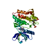
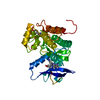
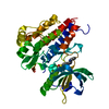
- Structure visualization
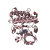
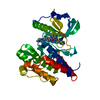
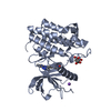
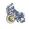
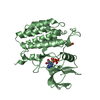
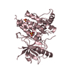
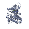
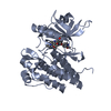
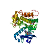
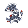
| Structure viewer | Molecule: MolmilJmol/JSmol |
|---|
- Downloads & links
-Download
| PDBx/mmCIF format | 4yjr.cif.gz | 228.4 KB | Display | PDBx/mmCIF format |
|---|---|---|---|---|
| PDB format | pdb4yjr.ent.gz | 184.4 KB | Display | PDB format |
| PDBx/mmJSON format | 4yjr.json.gz | Tree view | PDBx/mmJSON format | |
| Others |  Other downloads Other downloads |
-Validation report
| Arichive directory | https://data.pdbj.org/pub/pdb/validation_reports/yj/4yjrftp://data.pdbj.org/pub/pdb/validation_reports/yj/4yjr | HTTPS FTP |
|---|
-Related structure data
| Related structure data |  3srvC  4yjoC  4yjpC  4yjqC  4yjsC  4yjtC  4yjuC C: citing same article ( |
|---|---|
| Similar structure data |


-Links
 PDBj
PDBj


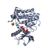
- Assembly
| Deposited unit | 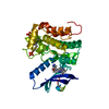
| ||||||||
|---|---|---|---|---|---|---|---|---|---|
| 1 |
| ||||||||
| Unit cell |
|
-Components
| #1: Protein | Mass: 32810.605 Da / Num. of mol.: 1 / Fragment: UNP residues 355-635 Source method: isolated from a genetically manipulated source Source: (gene. exp.) Homo sapiens (human) / Gene: SYK / Cell line (production host): Sf9 / Production host:   Spodoptera frugiperda (fall armyworm) Spodoptera frugiperda (fall armyworm)References: UniProt: P43405, non-specific protein-tyrosine kinase |
|---|---|
| #2: Chemical | ChemComp-4DJ / 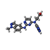  Mass: 414.463 Da / Num. of mol.: 1 / Source method: obtained synthetically / Formula: C22H22N8O Mass: 414.463 Da / Num. of mol.: 1 / Source method: obtained synthetically / Formula: C22H22N8O |
| #3: Water | ChemComp-HOH /  Mass: 18.015 Da / Num. of mol.: 347 / Source method: isolated from a natural source / Formula: H2O Mass: 18.015 Da / Num. of mol.: 347 / Source method: isolated from a natural source / Formula: H2O |
| Has protein modification | Y |
-Experimental details
-Experiment
| Experiment | Method: X-RAY DIFFRACTION / Number of used crystals: 1 |
|---|
- Sample preparation
| Crystal | Density Matthews: 2.1 Å3/Da / Density % sol: 41.5 % |
|---|---|
| Crystal grow | Temperature: 293 K / Method: vapor diffusion, hanging drop / pH: 6 Details: 30% Jeffamine ED-2001, 10% Glycerol, 0.1M MES pH6.0, 5mM TCEP |
-Data collection
| Diffraction | Mean temperature: 100 K | |||||||||||||||||||||||||||||||||||||||||||||||||||||||||||||||||||||||||||||||||||||||||||||||||||
|---|---|---|---|---|---|---|---|---|---|---|---|---|---|---|---|---|---|---|---|---|---|---|---|---|---|---|---|---|---|---|---|---|---|---|---|---|---|---|---|---|---|---|---|---|---|---|---|---|---|---|---|---|---|---|---|---|---|---|---|---|---|---|---|---|---|---|---|---|---|---|---|---|---|---|---|---|---|---|---|---|---|---|---|---|---|---|---|---|---|---|---|---|---|---|---|---|---|---|---|---|
| Diffraction source | Source: SYNCHROTRON / Site: ESRF  / Beamline: ID14-2 / Wavelength: 0.933 Å / Beamline: ID14-2 / Wavelength: 0.933 Å | |||||||||||||||||||||||||||||||||||||||||||||||||||||||||||||||||||||||||||||||||||||||||||||||||||
| Detector | Type: ADSC QUANTUM 4 / Detector: CCD / Date: Apr 28, 2005 | |||||||||||||||||||||||||||||||||||||||||||||||||||||||||||||||||||||||||||||||||||||||||||||||||||
| Radiation | Protocol: SINGLE WAVELENGTH / Monochromatic (M) / Laue (L): M / Scattering type: x-ray | |||||||||||||||||||||||||||||||||||||||||||||||||||||||||||||||||||||||||||||||||||||||||||||||||||
| Radiation wavelength | Wavelength: 0.933 Å / Relative weight: 1 | |||||||||||||||||||||||||||||||||||||||||||||||||||||||||||||||||||||||||||||||||||||||||||||||||||
| Reflection | Resolution: 1.25→29.128 Å / Num. obs: 74248 / % possible obs: 99.8 % / Redundancy: 3.6 % / Biso Wilson estimate: 13.3 Å2 / Rmerge(I) obs: 0.049 / Rsym value: 0.049 / Net I/av σ(I): 7.984 / Net I/σ(I): 13.8 / Num. measured all: 267412 | |||||||||||||||||||||||||||||||||||||||||||||||||||||||||||||||||||||||||||||||||||||||||||||||||||
| Reflection shell | Diffraction-ID: 1 / Rejects: _
|
- Processing
| Software |
| |||||||||||||||||||||||||||||||||||||||||||||||||||||||||||||||||||||||||||||||||||||||||||||||||||||||||||||||||||||||||||||
|---|---|---|---|---|---|---|---|---|---|---|---|---|---|---|---|---|---|---|---|---|---|---|---|---|---|---|---|---|---|---|---|---|---|---|---|---|---|---|---|---|---|---|---|---|---|---|---|---|---|---|---|---|---|---|---|---|---|---|---|---|---|---|---|---|---|---|---|---|---|---|---|---|---|---|---|---|---|---|---|---|---|---|---|---|---|---|---|---|---|---|---|---|---|---|---|---|---|---|---|---|---|---|---|---|---|---|---|---|---|---|---|---|---|---|---|---|---|---|---|---|---|---|---|---|---|---|
| Refinement | Method to determine structure: FOURIER SYNTHESIS Starting model: In-house SYK model Resolution: 1.32→29 Å / Cor.coef. Fo:Fc: 0.966 / Cor.coef. Fo:Fc free: 0.9498 / SU R Cruickshank DPI: 0.05 / Cross valid method: THROUGHOUT / σ(F): 0 / SU R Blow DPI: 0.046 / SU Rfree Blow DPI: 0.048 / SU Rfree Cruickshank DPI: 0.048
| |||||||||||||||||||||||||||||||||||||||||||||||||||||||||||||||||||||||||||||||||||||||||||||||||||||||||||||||||||||||||||||
| Displacement parameters | Biso max: 104.77 Å2 / Biso mean: 18.53 Å2 / Biso min: 3 Å2
| |||||||||||||||||||||||||||||||||||||||||||||||||||||||||||||||||||||||||||||||||||||||||||||||||||||||||||||||||||||||||||||
| Refine analyze | Luzzati coordinate error obs: 0.146 Å | |||||||||||||||||||||||||||||||||||||||||||||||||||||||||||||||||||||||||||||||||||||||||||||||||||||||||||||||||||||||||||||
| Refinement step | Cycle: final / Resolution: 1.32→29 Å
| |||||||||||||||||||||||||||||||||||||||||||||||||||||||||||||||||||||||||||||||||||||||||||||||||||||||||||||||||||||||||||||
| LS refinement shell | Resolution: 1.32→1.35 Å / Total num. of bins used: 20
| |||||||||||||||||||||||||||||||||||||||||||||||||||||||||||||||||||||||||||||||||||||||||||||||||||||||||||||||||||||||||||||
| Refinement TLS params. | Method: refined / Refine-ID: X-RAY DIFFRACTION
| |||||||||||||||||||||||||||||||||||||||||||||||||||||||||||||||||||||||||||||||||||||||||||||||||||||||||||||||||||||||||||||
| Refinement TLS group |
|
