 Movie
Movie Controller


+ Open data

- Basic information
| Entry | Database: PDB / ID: 6y23 | ||||||
|---|---|---|---|---|---|---|---|
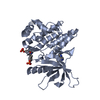
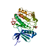
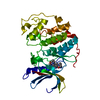
| Title | DDR1 kinase autoinhibited by its juxtamembrane region | ||||||
 Components Components | Epithelial discoidin domain-containing receptor 1 | ||||||
 Keywords Keywords | TRANSFERASE / Kinase / Collagen / Cell Signalling | ||||||
| Function / homology |  Function and homology information Function and homology informationprotein tyrosine kinase collagen receptor activity / smooth muscle cell-matrix adhesion / collagen-activated tyrosine kinase receptor signaling pathway / regulation of extracellular matrix disassembly / regulation of cell-matrix adhesion / ear development / branching involved in mammary gland duct morphogenesis / peptidyl-tyrosine autophosphorylation / wound healing, spreading of cells / neuron projection extension ...protein tyrosine kinase collagen receptor activity / smooth muscle cell-matrix adhesion / collagen-activated tyrosine kinase receptor signaling pathway / regulation of extracellular matrix disassembly / regulation of cell-matrix adhesion / ear development / branching involved in mammary gland duct morphogenesis / peptidyl-tyrosine autophosphorylation / wound healing, spreading of cells / neuron projection extension / smooth muscle cell migration / axon development / mammary gland alveolus development / Non-integrin membrane-ECM interactions / collagen binding / embryo implantation / lactation / transmembrane receptor protein tyrosine kinase activity / cell surface receptor protein tyrosine kinase signaling pathway / regulation of cell growth / positive regulation of neuron projection development / receptor protein-tyrosine kinase / protein autophosphorylation / positive regulation of phosphatidylinositol 3-kinase/protein kinase B signal transduction / cell population proliferation / cell adhesion / signaling receptor complex / negative regulation of cell population proliferation / : / extracellular exosome / ATP binding / metal ion binding / plasma membrane Similarity search - Function | ||||||
| Biological species |  Homo sapiens (human) Homo sapiens (human) | ||||||
| Method |  X-RAY DIFFRACTION / SYNCHROTRON / MOLECULAR REPLACEMENT / Resolution: 2.58 Å X-RAY DIFFRACTION / SYNCHROTRON / MOLECULAR REPLACEMENT / Resolution: 2.58 Å | ||||||
 Authors Authors | Sammon, D. / Hohenester, E. / Leitinger, B. | ||||||
| Funding support |  United Kingdom, 1items United Kingdom, 1items
| ||||||
 Citation Citation | Journal: Proc.Natl.Acad.Sci.USA / Year: 2020 Title: Two-step release of kinase autoinhibition in discoidin domain receptor 1. Authors: Sammon, D. / Hohenester, E. / Leitinger, B. | ||||||
| History |
|
- Structure visualization
| Structure viewer | Molecule: MolmilJmol/JSmol |
|---|
- Downloads & links
-Download
| PDBx/mmCIF format | 6y23.cif.gz | 532.6 KB | Display | PDBx/mmCIF format |
|---|---|---|---|---|
| PDB format | pdb6y23.ent.gz | 430.6 KB | Display | PDB format |
| PDBx/mmJSON format | 6y23.json.gz | Tree view | PDBx/mmJSON format | |
| Others |  Other downloads Other downloads |
-Validation report
| Arichive directory | https://data.pdbj.org/pub/pdb/validation_reports/y2/6y23ftp://data.pdbj.org/pub/pdb/validation_reports/y2/6y23 | HTTPS FTP |
|---|
-Related structure data
| Related structure data |  5bvwS S: Starting model for refinement |
|---|---|
| Similar structure data |


-Links
 PDBj
PDBj


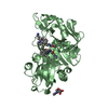
- Assembly
| Deposited unit | 
| ||||||||||||
|---|---|---|---|---|---|---|---|---|---|---|---|---|---|
| 1 | 
| ||||||||||||
| 2 | 
| ||||||||||||
| 3 | 
| ||||||||||||
| Unit cell |
|
-Components
| #1: Protein | Mass: 39045.703 Da / Num. of mol.: 3 Source method: isolated from a genetically manipulated source Details: Vector derived GP at N-terminus, Y569F and Y586F mutations Source: (gene. exp.) Homo sapiens (human) / Gene: DDR1, CAK, EDDR1, NEP, NTRK4, PTK3A, RTK6, TRKE / Production host:   Spodoptera frugiperda (fall armyworm) Spodoptera frugiperda (fall armyworm)References: UniProt: Q08345, receptor protein-tyrosine kinase #2: Chemical | ChemComp-SO4 /   Mass: 96.063 Da / Num. of mol.: 8 / Source method: obtained synthetically / Formula: SO4 Mass: 96.063 Da / Num. of mol.: 8 / Source method: obtained synthetically / Formula: SO4#3: Water | ChemComp-HOH / |  Mass: 18.015 Da / Num. of mol.: 40 / Source method: isolated from a natural source / Formula: H2O Mass: 18.015 Da / Num. of mol.: 40 / Source method: isolated from a natural source / Formula: H2OHas ligand of interest | N | |
|---|
-Experimental details
-Experiment
| Experiment | Method: X-RAY DIFFRACTION / Number of used crystals: 1 |
|---|
- Sample preparation
| Crystal | Density Matthews: 2.78 Å3/Da / Density % sol: 55.76 % |
|---|---|
| Crystal grow | Temperature: 277 K / Method: vapor diffusion, sitting drop Details: 9.5 mg/ml protein in 20 mM HEPES pH 7.5, 200 mM NaCl, 1 mM TCEP. Precipitant: 100 mM Tris pH 8.0, 1.5 M ammonium sulphate. |
-Data collection
| Diffraction | Mean temperature: 100 K / Serial crystal experiment: N |
|---|---|
| Diffraction source | Source: SYNCHROTRON / Site: Diamond / Beamline: I24 / Wavelength: 0.9686 Å |
| Detector | Type: DECTRIS PILATUS3 6M / Detector: PIXEL / Date: Oct 5, 2018 |
| Radiation | Protocol: SINGLE WAVELENGTH / Monochromatic (M) / Laue (L): M / Scattering type: x-ray |
| Radiation wavelength | Wavelength: 0.9686 Å / Relative weight: 1 |
| Reflection | Resolution: 2.58→58.13 Å / Num. obs: 43468 / % possible obs: 100 % / Redundancy: 20.3 % / Biso Wilson estimate: 41.5 Å2 / CC1/2: 0.979 / Rpim(I) all: 0.128 / Net I/σ(I): 6.5 |
| Reflection shell | Resolution: 2.58→2.62 Å / Redundancy: 20.9 % / Mean I/σ(I) obs: 1.2 / Num. unique obs: 2087 / CC1/2: 0.443 / Rpim(I) all: 1.129 / % possible all: 99.1 |
- Processing
| Software |
| |||||||||||||||||||||||||||||||||||||||||||||||||||||||||||||||||||||||||||||||||||||||||||||||||||||||||||||||||||||||
|---|---|---|---|---|---|---|---|---|---|---|---|---|---|---|---|---|---|---|---|---|---|---|---|---|---|---|---|---|---|---|---|---|---|---|---|---|---|---|---|---|---|---|---|---|---|---|---|---|---|---|---|---|---|---|---|---|---|---|---|---|---|---|---|---|---|---|---|---|---|---|---|---|---|---|---|---|---|---|---|---|---|---|---|---|---|---|---|---|---|---|---|---|---|---|---|---|---|---|---|---|---|---|---|---|---|---|---|---|---|---|---|---|---|---|---|---|---|---|---|---|
| Refinement | Method to determine structure: MOLECULAR REPLACEMENT Starting model: 5bvw Resolution: 2.58→54.42 Å / SU ML: 0.34 / Cross valid method: FREE R-VALUE / σ(F): 1.34 / Phase error: 26.7489
| |||||||||||||||||||||||||||||||||||||||||||||||||||||||||||||||||||||||||||||||||||||||||||||||||||||||||||||||||||||||
| Solvent computation | Shrinkage radii: 0.9 Å / VDW probe radii: 1.11 Å | |||||||||||||||||||||||||||||||||||||||||||||||||||||||||||||||||||||||||||||||||||||||||||||||||||||||||||||||||||||||
| Displacement parameters | Biso mean: 47.65 Å2 | |||||||||||||||||||||||||||||||||||||||||||||||||||||||||||||||||||||||||||||||||||||||||||||||||||||||||||||||||||||||
| Refinement step | Cycle: LAST / Resolution: 2.58→54.42 Å
| |||||||||||||||||||||||||||||||||||||||||||||||||||||||||||||||||||||||||||||||||||||||||||||||||||||||||||||||||||||||
| Refine LS restraints |
| |||||||||||||||||||||||||||||||||||||||||||||||||||||||||||||||||||||||||||||||||||||||||||||||||||||||||||||||||||||||
| LS refinement shell |
|
