[English] 日本語
Yorodumi
- PDB-3qc4: PDK1 in complex with DFG-OUT inhibitor xxx -

+
Open data


ID or keywords:

Loading...

-
Basic information

Entry
Database: PDB / ID: 3qc4
TitlePDK1 in complex with DFG-OUT inhibitor xxx
Components3-phosphoinositide-dependent protein kinase 1
KeywordsTRANSFERASE/TRANSFERASE INHIBITOR / serine/threonine kinase / phosphorylation / TRANSFERASE-TRANSFERASE INHIBITOR complex
Function / homology
Function and homology information


3-phosphoinositide-dependent protein kinase activity / Activation of AKT2 / regulation of mast cell degranulation / negative regulation of toll-like receptor signaling pathway / type B pancreatic cell development / positive regulation of phospholipase activity / RSK activation / hyperosmotic response / regulation of canonical NF-kappaB signal transduction / positive regulation of vascular endothelial cell proliferation ...3-phosphoinositide-dependent protein kinase activity / Activation of AKT2 / regulation of mast cell degranulation / negative regulation of toll-like receptor signaling pathway / type B pancreatic cell development / positive regulation of phospholipase activity / RSK activation / hyperosmotic response / regulation of canonical NF-kappaB signal transduction / positive regulation of vascular endothelial cell proliferation / negative regulation of cardiac muscle cell apoptotic process / phospholipase activator activity / positive regulation of sprouting angiogenesis / Constitutive Signaling by AKT1 E17K in Cancer / CD28 dependent PI3K/Akt signaling / phospholipase binding / positive regulation of blood vessel endothelial cell migration / Role of LAT2/NTAL/LAB on calcium mobilization / Estrogen-stimulated signaling through PRKCZ / SARS-CoV-2 targets host intracellular signalling and regulatory pathways / negative regulation of endothelial cell apoptotic process / SARS-CoV-1 targets host intracellular signalling and regulatory pathways / extrinsic apoptotic signaling pathway / RHO GTPases activate PKNs / cellular response to epidermal growth factor stimulus / GPVI-mediated activation cascade / T cell costimulation / activation of protein kinase B activity / Integrin signaling / insulin-like growth factor receptor signaling pathway / positive regulation of release of sequestered calcium ion into cytosol / VEGFR2 mediated vascular permeability / VEGFR2 mediated cell proliferation / cell projection / calcium-mediated signaling / positive regulation of protein localization to plasma membrane / peptidyl-threonine phosphorylation / negative regulation of transforming growth factor beta receptor signaling pathway / negative regulation of protein kinase activity / epidermal growth factor receptor signaling pathway / CLEC7A (Dectin-1) signaling / FCERI mediated NF-kB activation / G beta:gamma signalling through PI3Kgamma / cellular response to insulin stimulus / positive regulation of angiogenesis / Regulation of TP53 Degradation / cell migration / Downstream TCR signaling / insulin receptor signaling pathway / PIP3 activates AKT signaling / cytoplasmic vesicle / actin cytoskeleton organization / protein autophosphorylation / postsynaptic density / positive regulation of phosphatidylinositol 3-kinase/protein kinase B signal transduction / non-specific serine/threonine protein kinase / intracellular signal transduction / protein phosphorylation / protein serine kinase activity / focal adhesion / protein serine/threonine kinase activity / ATP binding / nucleus / plasma membrane / cytoplasm / cytosol
Similarity search - Function
PDK1-type, PH domain / PDPK1 family / PH domain / PH-like domain superfamily / Transferase(Phosphotransferase) domain 1 / Transferase(Phosphotransferase); domain 1 / Phosphorylase Kinase; domain 1 / Phosphorylase Kinase; domain 1 / Serine/threonine-protein kinase, active site / Serine/Threonine protein kinases active-site signature. ...PDK1-type, PH domain / PDPK1 family / PH domain / PH-like domain superfamily / Transferase(Phosphotransferase) domain 1 / Transferase(Phosphotransferase); domain 1 / Phosphorylase Kinase; domain 1 / Phosphorylase Kinase; domain 1 / Serine/threonine-protein kinase, active site / Serine/Threonine protein kinases active-site signature. / Protein kinase domain / Serine/Threonine protein kinases, catalytic domain / Protein kinase, ATP binding site / Protein kinases ATP-binding region signature. / Protein kinase domain profile. / Protein kinase domain / Protein kinase-like domain superfamily / 2-Layer Sandwich / Orthogonal Bundle / Mainly Alpha / Alpha Beta
Similarity search - Domain/homology
Chem-MP7 / 3-phosphoinositide-dependent protein kinase 1
Similarity search - Component
Biological speciesHomo sapiens (human)
MethodX-RAY DIFFRACTION / SYNCHROTRON / MOLECULAR REPLACEMENT / Resolution: 1.8 Å
AuthorsArndt, J.W.
CitationJournal: Bioorg.Med.Chem.Lett. / Year: 2011
Title: Discovery of a potent and highly selective PDK1 inhibitor via fragment-based drug discovery.
Authors: Erlanson, D.A. / Arndt, J.W. / Cancilla, M.T. / Cao, K. / Elling, R.A. / English, N. / Friedman, J. / Hansen, S.K. / Hession, C. / Joseph, I. / Kumaravel, G. / Lee, W.C. / Lind, K.E. / ...Authors: Erlanson, D.A. / Arndt, J.W. / Cancilla, M.T. / Cao, K. / Elling, R.A. / English, N. / Friedman, J. / Hansen, S.K. / Hession, C. / Joseph, I. / Kumaravel, G. / Lee, W.C. / Lind, K.E. / McDowell, R.S. / Miatkowski, K. / Nguyen, C. / Nguyen, T.B. / Park, S. / Pathan, N. / Penny, D.M. / Romanowski, M.J. / Scott, D. / Silvian, L. / Simmons, R.L. / Tangonan, B.T. / Yang, W. / Sun, L.
History
DepositionJan 15, 2011Deposition site: RCSB / Processing site: RCSB
Revision 1.0Apr 20, 2011Provider: repository / Type: Initial release
Revision 1.1Jul 13, 2011Group: Version format compliance

-
Structure visualization

Structure viewerMolecule:
MolmilJmol/JSmol

Downloads & links

-
Assembly

Deposited unit
A: 3-phosphoinositide-dependent protein kinase 1
B: 3-phosphoinositide-dependent protein kinase 1
hetero molecules


Theoretical massNumber of molelcules
Total (without water)72,4794
Polymers71,4462
Non-polymers1,0332
Water7,656425
1
A: 3-phosphoinositide-dependent protein kinase 1
hetero molecules


Theoretical massNumber of molelcules
Total (without water)36,2392
Polymers35,7231
Non-polymers5161
Water181
TypeNameSymmetry operationNumber
identity operation1_555x,y,z1
2
B: 3-phosphoinositide-dependent protein kinase 1
hetero molecules


Theoretical massNumber of molelcules
Total (without water)36,2392
Polymers35,7231
Non-polymers5161
Water181
TypeNameSymmetry operationNumber
identity operation1_555x,y,z1
Unit cell
Length a, b, c (Å)183.147, 57.336, 83.694
Angle α, β, γ (deg.)90.00, 114.13, 90.00
Int Tables number5
Space group name H-MC121

-
Components

#1: Protein 3-phosphoinositide-dependent protein kinase 1 / hPDK1


Mass: 35722.922 Da / Num. of mol.: 2 / Fragment: kinase domain (UNP residues 51-359) / Mutation: E303A,K304A
Source method: isolated from a genetically manipulated source
Source: (gene. exp.) Homo sapiens (human) / Gene: PDPK1, PDK1 / Production host: Spodoptera frugiperda (fall armyworm)
References: UniProt: O15530, non-specific serine/threonine protein kinase
#2: Chemical ChemComp-MP7 / 1-(3,4-difluorobenzyl)-2-oxo-N-{(1R)-2-[(2-oxo-2,3-dihydro-1H-benzimidazol-5-yl)oxy]-1-phenylethyl}-1,2-dihydropyridine-3-carboxamide


Mass: 516.495 Da / Num. of mol.: 2 / Source method: obtained synthetically / Formula: C28H22F2N4O4
#3: Water ChemComp-HOH / water


Mass: 18.015 Da / Num. of mol.: 425 / Source method: isolated from a natural source / Formula: H2O
Nonpolymer detailsLIGAND MP7 NAMED COMPOUND 33 IN PRIMARY CITATION.

-
Experimental details

-
Experiment

ExperimentMethod: X-RAY DIFFRACTION / Number of used crystals: 1

-
Sample preparation

CrystalDensity Matthews: 2.81 Å3/Da / Density % sol: 56.17 %
Crystal growTemperature: 293 K / Method: vapor diffusion, sitting drop / pH: 7
Details: 2 M ammonium sulfate, pH 7, VAPOR DIFFUSION, SITTING DROP, temperature 293K

-
Data collection

DiffractionMean temperature: 100 K
Diffraction sourceSource: SYNCHROTRON / Site: APS / Beamline: 31-ID / Wavelength: 0.97
DetectorType: RAYONIX MX225HE / Detector: CCD / Date: Aug 13, 2010
RadiationMonochromator: Kohzu HLD-4 Double Crystal / Protocol: SINGLE WAVELENGTH / Monochromatic (M) / Laue (L): M / Scattering type: x-ray
Radiation wavelengthWavelength: 0.97 Å / Relative weight: 1
ReflectionResolution: 1.8→50 Å / Num. obs: 72110 / % possible obs: 98.3 % / Observed criterion σ(F): 2 / Observed criterion σ(I): 2 / Redundancy: 4 % / Rmerge(I) obs: 0.065
Reflection shellResolution: 1.8→1.86 Å / % possible all: 97.8

-
Processing

Software
NameVersionClassification
CrystalCleardata collection
MOLREPphasing
REFMAC5.5.0109refinement
HKL-2000data reduction
HKL-2000data scaling
Refinement
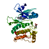
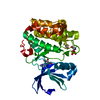
Method to determine structure: MOLECULAR REPLACEMENT / Resolution: 1.8→30.49 Å / Cor.coef. Fo:Fc: 0.964 / Cor.coef. Fo:Fc free: 0.947 / SU B: 5.087 / SU ML: 0.071 / Cross valid method: THROUGHOUT / σ(F): 2 / ESU R: 0.102 / ESU R Free: 0.106 / Stereochemistry target values: MAXIMUM LIKELIHOOD
Details: HYDROGENS HAVE BEEN ADDED IN THE RIDING POSITIONS U VALUES: RESIDUAL ONLY
RfactorNum. reflection% reflectionSelection details
Rfree0.21321 3620 5 %RANDOM
Rwork0.17239 ---
obs0.17446 68387 98.04 %-
all-73398 --
Solvent computationIon probe radii: 0.8 Å / Shrinkage radii: 0.8 Å / VDW probe radii: 1.4 Å / Solvent model: MASK
Displacement parametersBiso mean: 40.295 Å2
Baniso -1Baniso -2Baniso -3
1--1.23 Å20 Å2-1.16 Å2
2--2.1 Å20 Å2
3----1.81 Å2
Refinement stepCycle: LAST / Resolution: 1.8→30.49 Å
ProteinNucleic acidLigandSolventTotal
Num. atoms4489 0 76 425 4990
Refine LS restraints
Refine-IDTypeDev idealDev ideal targetNumber
X-RAY DIFFRACTIONr_bond_refined_d0.030.0224697
X-RAY DIFFRACTIONr_bond_other_d
X-RAY DIFFRACTIONr_angle_refined_deg2.2881.9976353
X-RAY DIFFRACTIONr_angle_other_deg
X-RAY DIFFRACTIONr_dihedral_angle_1_deg6.3485558
X-RAY DIFFRACTIONr_dihedral_angle_2_deg34.823.78209
X-RAY DIFFRACTIONr_dihedral_angle_3_deg16.03215.036825
X-RAY DIFFRACTIONr_dihedral_angle_4_deg20.8291524
X-RAY DIFFRACTIONr_chiral_restr0.1910.2686
X-RAY DIFFRACTIONr_gen_planes_refined0.0140.0213528
X-RAY DIFFRACTIONr_gen_planes_other
X-RAY DIFFRACTIONr_nbd_refined
X-RAY DIFFRACTIONr_nbd_other
X-RAY DIFFRACTIONr_nbtor_refined
X-RAY DIFFRACTIONr_nbtor_other
X-RAY DIFFRACTIONr_xyhbond_nbd_refined
X-RAY DIFFRACTIONr_xyhbond_nbd_other
X-RAY DIFFRACTIONr_metal_ion_refined
X-RAY DIFFRACTIONr_metal_ion_other
X-RAY DIFFRACTIONr_symmetry_vdw_refined
X-RAY DIFFRACTIONr_symmetry_vdw_other
X-RAY DIFFRACTIONr_symmetry_hbond_refined
X-RAY DIFFRACTIONr_symmetry_hbond_other
X-RAY DIFFRACTIONr_symmetry_metal_ion_refined
X-RAY DIFFRACTIONr_symmetry_metal_ion_other
X-RAY DIFFRACTIONr_mcbond_it1.4471.52779
X-RAY DIFFRACTIONr_mcbond_other
X-RAY DIFFRACTIONr_mcangle_it2.40424484
X-RAY DIFFRACTIONr_scbond_it3.64231918
X-RAY DIFFRACTIONr_scangle_it5.6124.51865
X-RAY DIFFRACTIONr_rigid_bond_restr
X-RAY DIFFRACTIONr_sphericity_free
X-RAY DIFFRACTIONr_sphericity_bonded
LS refinement shellResolution: 1.802→1.849 Å / Total num. of bins used: 20
RfactorNum. reflection% reflection
Rfree0.291 244 -
Rwork0.253 4973 -
obs--96.25 %
Refinement TLS params.

Method: refined / Refine-ID: X-RAY DIFFRACTION

IDL112)L122)L132)L222)L232)L332)S11 (Å °)S12 (Å °)S13 (Å °)S21 (Å °)S22 (Å °)S23 (Å °)S31 (Å °)S32 (Å °)S33 (Å °)T112)T122)T132)T222)T232)T332)Origin x (Å)Origin y (Å)Origin z (Å)
19.3803-0.69713.05215.45280.91973.3057-0.4721-0.1371.1811-0.09620.5566-1.3252-0.02070.2031-0.08450.0693-0.00110.00540.1673-0.18540.53618.864938.525921.9757
20.84470.0532-0.14520.53320.16831.84280.0190.029-0.0107-0.0450.0441-0.0690.11510.1425-0.06310.07140.03610.02040.0902-0.01360.0435-3.182824.123722.2782
36.2711-1.82892.84684.87225.557110.74570.06360.2366-0.0168-0.22280.3831-0.3496-0.33790.7012-0.44660.1124-0.01490.00280.1678-0.0170.15027.12689.51945.851
413.4829-1.2221-1.61428.06580.57132.1730.14242.52231.8029-0.8254-0.3065-0.7917-0.07490.15740.16420.1752-0.00750.15650.62590.44430.489118.997254.927745.2782
57.3132-1.93981.99364.3852-1.23782.6782-0.1061.02821.2257-0.3555-0.2555-0.3986-0.32130.18280.36150.1926-0.0302-0.03070.16090.18810.31649.746955.681751.5559
60.93080.31680.5591.0820.26791.44970.03620.02890.05130.0665-0.0479-0.11370.02170.04260.01170.05750.0205-0.02470.03140.00130.07584.795535.102661.8392
716.30442.76260.09121.4899-3.08829.4311-0.2539-0.51170.5311-0.0683-0.2833-0.11720.10130.35470.53720.0578-0.00340.09020.2592-0.12010.251712.55133.199121.8765
87.47154.08084.71232.75081.85333.9965-0.1390.06760.1805-0.1515-0.1237-0.02230.00840.2030.26280.1493-0.01170.00270.18970.11380.17129.648549.254251.9179
Refinement TLS group
IDRefine-IDRefine TLS-IDAuth asym-IDAuth seq-ID
1X-RAY DIFFRACTION1A76 - 163
2X-RAY DIFFRACTION2A164 - 359
3X-RAY DIFFRACTION3B51 - 55
4X-RAY DIFFRACTION4B76 - 118
5X-RAY DIFFRACTION5B127 - 163
6X-RAY DIFFRACTION6B164 - 359
7X-RAY DIFFRACTION7A1
8X-RAY DIFFRACTION8B2

+
About Yorodumi

-
News

-
Feb 9, 2022. New format data for meta-information of EMDB entries

New format data for meta-information of EMDB entries

  • Version 3 of the EMDB header file is now the official format.
  • The previous official version 1.9 will be removed from the archive.

Related info.:EMDB header

External links:wwPDB to switch to version 3 of the EMDB data model

-
Aug 12, 2020. Covid-19 info

Covid-19 info

URL: https://pdbjlvh1.pdbj.org/emnavi/covid19.php

New page: Covid-19 featured information page in EM Navigator.

Related info.:Covid-19 info / Mar 5, 2020. Novel coronavirus structure data

+
Mar 5, 2020. Novel coronavirus structure data

Novel coronavirus structure data

Related info.:Yorodumi Speices / Aug 12, 2020. Covid-19 info

External links:COVID-19 featured content - PDBj / Molecule of the Month (242):Coronavirus Proteases

+
Jan 31, 2019. EMDB accession codes are about to change! (news from PDBe EMDB page)

EMDB accession codes are about to change! (news from PDBe EMDB page)

  • The allocation of 4 digits for EMDB accession codes will soon come to an end. Whilst these codes will remain in use, new EMDB accession codes will include an additional digit and will expand incrementally as the available range of codes is exhausted. The current 4-digit format prefixed with “EMD-” (i.e. EMD-XXXX) will advance to a 5-digit format (i.e. EMD-XXXXX), and so on. It is currently estimated that the 4-digit codes will be depleted around Spring 2019, at which point the 5-digit format will come into force.
  • The EM Navigator/Yorodumi systems omit the EMD- prefix.

Related info.:Q: What is EMD? / ID/Accession-code notation in Yorodumi/EM Navigator

External links:EMDB Accession Codes are Changing Soon! / Contact to PDBj

+
Jul 12, 2017. Major update of PDB

Major update of PDB

  • wwPDB released updated PDB data conforming to the new PDBx/mmCIF dictionary.
  • This is a major update changing the version number from 4 to 5, and with Remediation, in which all the entries are updated.
  • In this update, many items about electron microscopy experimental information are reorganized (e.g. em_software).
  • Now, EM Navigator and Yorodumi are based on the updated data.

External links:wwPDB Remediation / Enriched Model Files Conforming to OneDep Data Standards Now Available in the PDB FTP Archive

-
Yorodumi

Thousand views of thousand structures

  • Yorodumi is a browser for structure data from EMDB, PDB, SASBDB, etc.
  • This page is also the successor to EM Navigator detail page, and also detail information page/front-end page for Omokage search.
  • The word "yorodu" (or yorozu) is an old Japanese word meaning "ten thousand". "mi" (miru) is to see.

Related info.:EMDB / PDB / SASBDB / Comparison of 3 databanks / Yorodumi Search / Aug 31, 2016. New EM Navigator & Yorodumi / Yorodumi Papers / Jmol/JSmol / Function and homology information / Changes in new EM Navigator and Yorodumi

Read more