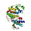
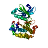
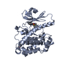
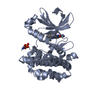
Entry Database : PDB / ID : 3nayTitle PDK1 in complex with inhibitor MP6 3-phosphoinositide-dependent protein kinase 1 Keywords / / Function / homology Function Domain/homology Component
/ / / / / / / / / / / / / / / / / / / / / / / / / / / / / / / / / / / / / / / / / / / / / / / / / / / / / / / / / / / / / / / / / / / / / / / / / / / / / / / / / / / / / / / / / / / / / / / / / / / / / / / / / Biological species Homo sapiens (human)Method / / / Resolution : 2.6 Å Authors Yan, Y. / Munshi, S.K. / Allison, T. Journal : J.Biol.Chem. / Year : 2010Title : Selective inhibition of PDK1 using an allosteric kinase inhibitor and RNAi impairs cancer cell migration and anchorage-independent growth of primary tumor linesAuthors: Nagashima, K. / Shumway, S.D. / Sathyanarayanan, S. / Chen, A. / Dolinski, B. / Xu, Y. / Keilhack, H. / Nguyen, T. / Wiznerowicz, M. / Li, L. / Lutterbach, B.A. / Paweletz, C. / Allison, T. ... Authors : Nagashima, K. / Shumway, S.D. / Sathyanarayanan, S. / Chen, A. / Dolinski, B. / Xu, Y. / Keilhack, H. / Nguyen, T. / Wiznerowicz, M. / Li, L. / Lutterbach, B.A. / Paweletz, C. / Allison, T. / Yan, Y. / Munshi, S.K. / Klippel, A. / Kraus, M. / Bobkova, E.V. / Deshmukh, S. / Xu, Z. / Mueller, U. / Szewczak, A.A. / Pan, B.-S. / Richon, V. / Pollock, R. / Blume-Jensen, P. / Northrup, A. / Andersen, J.N. History Deposition Jun 2, 2010 Deposition site / Processing site Revision 1.0 Nov 24, 2010 Provider / Type Revision 1.1 Jul 13, 2011 Group Revision 1.2 Jan 24, 2018 Group / Category / Item Revision 1.3 Nov 6, 2024 Group Data collection / Database references ... Data collection / Database references / Derived calculations / Structure summary Category chem_comp_atom / chem_comp_bond ... chem_comp_atom / chem_comp_bond / database_2 / pdbx_entry_details / pdbx_modification_feature / struct_conn / struct_ref_seq_dif / struct_site Item _database_2.pdbx_DOI / _database_2.pdbx_database_accession ... _database_2.pdbx_DOI / _database_2.pdbx_database_accession / _struct_conn.pdbx_leaving_atom_flag / _struct_ref_seq_dif.details / _struct_site.pdbx_auth_asym_id / _struct_site.pdbx_auth_comp_id / _struct_site.pdbx_auth_seq_id
Show all Show less
 Movie
Movie Controller


 Open data
 Basic information
 Components
 Keywords
 Function and homology information
 Homo sapiens (human)
 X-RAY DIFFRACTION /
  Authors
 Citation
 Structure visualization
 Downloads & links
 Other downloads


 PDBj
PDBj


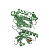
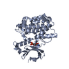
 Assembly


 Spodoptera frugiperda (fall armyworm) / Strain (production host): sf21
 Mass: 427.541 Da / Num. of mol.: 2 / Source method: obtained synthetically / Formula: C26H29N5O
 Mass: 18.015 Da / Num. of mol.: 149 / Source method: isolated from a natural source / Formula: H2O
 Sample preparation
 / Beamline: 17-BM / Wavelength: 1 Å
 Processing