regulation of relaxation of cardiac muscle / regulation of cellular localization / negative regulation of sodium ion transmembrane transport / regulation of cardiac muscle cell action potential involved in regulation of contraction / calcium- and calmodulin-dependent protein kinase complex / regulation of cell communication by electrical coupling / Ca2+/calmodulin-dependent protein kinase / regulation of the force of heart contraction / Trafficking of AMPA receptors / endoplasmic reticulum calcium ion homeostasis ...regulation of relaxation of cardiac muscle / regulation of cellular localization / negative regulation of sodium ion transmembrane transport / regulation of cardiac muscle cell action potential involved in regulation of contraction / calcium- and calmodulin-dependent protein kinase complex / regulation of cell communication by electrical coupling / Ca2+/calmodulin-dependent protein kinase / regulation of the force of heart contraction / Trafficking of AMPA receptors / endoplasmic reticulum calcium ion homeostasis / sodium channel inhibitor activity / calcium/calmodulin-dependent protein kinase activity / Assembly and cell surface presentation of NMDA receptors / regulation of calcium ion transmembrane transport via high voltage-gated calcium channel / relaxation of cardiac muscle / cardiac muscle cell contraction / CaMK IV-mediated phosphorylation of CREB / positive regulation of cardiac muscle hypertrophy / regulation of membrane depolarization / regulation of cardiac muscle cell action potential / regulation of heart contraction / Phase 0 - rapid depolarisation / Negative regulation of NMDA receptor-mediated neuronal transmission / Unblocking of NMDA receptors, glutamate binding and activation / regulation of heart rate by cardiac conduction / regulation of neuronal synaptic plasticity / regulation of cell communication by electrical coupling involved in cardiac conduction / Ion transport by P-type ATPases / regulation of ryanodine-sensitive calcium-release channel activity / Long-term potentiation / HSF1-dependent transactivation / Regulation of MECP2 expression and activity / regulation of protein localization to plasma membrane / regulation of release of sequestered calcium ion into cytosol by sarcoplasmic reticulum / Ion homeostasis / titin binding / regulation of cardiac muscle contraction by regulation of the release of sequestered calcium ion / positive regulation of cardiac muscle cell apoptotic process / sarcoplasmic reticulum membrane / cellular response to calcium ion / Ras activation upon Ca2+ influx through NMDA receptor / regulation of cell growth / RAF activation / sarcolemma / Interferon gamma signaling / long-term synaptic potentiation / Signaling by RAF1 mutants / Signaling by moderate kinase activity BRAF mutants / Paradoxical activation of RAF signaling by kinase inactive BRAF / Signaling downstream of RAS mutants / endocytic vesicle membrane / Signaling by BRAF and RAF1 fusions / RAF/MAP kinase cascade / protein phosphorylation / transmembrane transporter binding / calmodulin binding / neuron projection / postsynaptic density / protein serine kinase activity / protein serine/threonine kinase activity / regulation of transcription by RNA polymerase II / protein homodimerization activity / nucleoplasm / ATP binding / membrane / identical protein binding / nucleus / cytosol / cytoplasm Similarity search - Function
Calcium/calmodulin-dependent protein kinase II, association-domain / Calcium/calmodulin dependent protein kinase II association domain / NTF2-like domain superfamily / Phosphorylase Kinase; domain 1 / Phosphorylase Kinase; domain 1 / Transferase(Phosphotransferase) domain 1 / Transferase(Phosphotransferase); domain 1 / Serine/threonine-protein kinase, active site / Serine/Threonine protein kinases active-site signature. / Protein kinase domain ...Calcium/calmodulin-dependent protein kinase II, association-domain / Calcium/calmodulin dependent protein kinase II association domain / NTF2-like domain superfamily / Phosphorylase Kinase; domain 1 / Phosphorylase Kinase; domain 1 / Transferase(Phosphotransferase) domain 1 / Transferase(Phosphotransferase); domain 1 / Serine/threonine-protein kinase, active site / Serine/Threonine protein kinases active-site signature. / Protein kinase domain / Serine/Threonine protein kinases, catalytic domain / Protein kinase, ATP binding site / Protein kinases ATP-binding region signature. / Protein kinase domain profile. / Protein kinase domain / Protein kinase-like domain superfamily / 2-Layer Sandwich / Orthogonal Bundle / Mainly Alpha / Alpha Beta Similarity search - Domain/homology
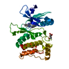
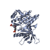
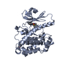
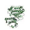
Chem-C2V / 2,3-DIHYDROXY-1,4-DITHIOBUTANE / Calcium/calmodulin-dependent protein kinase type II subunit delta Similarity search - Component
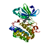
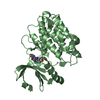
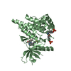
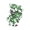
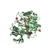
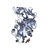
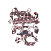
A: Calcium/calmodulin-dependent protein kinase type II subunit delta B: Calcium/calmodulin-dependent protein kinase type II subunit delta hetero molecules
Mass: 18.015 Da / Num. of mol.: 259 / Source method: isolated from a natural source / Formula: H2O
-
Experimental details
-
Experiment
Experiment
Method: X-RAY DIFFRACTION / Number of used crystals: 1
-
Sample preparation
Crystal
Density Matthews: 2.33 Å3/Da / Density % sol: 47.14 %
Crystal grow
Temperature: 293 K / Method: vapor diffusion, sitting drop Details: CamKII S3-K301, in 20mM imidazole pH 8.5, 0.3M sodium chloride, 5mM TCEP, was concentrated to 36 mg/ml and flash frozen in liquid nitrogen for long term storage at -80 C in 10 microL ...Details: CamKII S3-K301, in 20mM imidazole pH 8.5, 0.3M sodium chloride, 5mM TCEP, was concentrated to 36 mg/ml and flash frozen in liquid nitrogen for long term storage at -80 C in 10 microL aliquots. The protein was thawed and diluted down to 12 mg/mL in the same buffer just prior to crystallization experiments. Sitting drop vapor diffusion droplets were assembled with 250 nL of 12 mg/mL CamKII, 0.6 mM inhibitor and 250 nL of reservoir solution 24% peg 3350, 0.2 M ammonium tartrate, 0.1 M arginine. Flat crystal plates (typically 0.03 mm x 0.2 mm x 0.4 mm in size) grew in 4-7 days at 20 C. A crystal seed suspension was prepared with ten crushed crystals combined into 100 uL of reservoir solution and stored at -80 C. A thirty fold seed dilution was prepared in the same solution for addition to protein droplets in a 1 to 1 volume ratio to enhance crystallization of difficult to crystallize inhibitors.
-
Data collection
Diffraction
Mean temperature: 100 K
Diffraction source
Source: SYNCHROTRON / Site: ALS / Beamline: 5.0.2 / Wavelength: 1 Å
In the structure databanks used in Yorodumi, some data are registered as the other names, "COVID-19 virus" and "2019-nCoV". Here are the details of the virus and the list of structure data.
Jan 31, 2019. EMDB accession codes are about to change! (news from PDBe EMDB page)
EMDB accession codes are about to change! (news from PDBe EMDB page)
The allocation of 4 digits for EMDB accession codes will soon come to an end. Whilst these codes will remain in use, new EMDB accession codes will include an additional digit and will expand incrementally as the available range of codes is exhausted. The current 4-digit format prefixed with “EMD-” (i.e. EMD-XXXX) will advance to a 5-digit format (i.e. EMD-XXXXX), and so on. It is currently estimated that the 4-digit codes will be depleted around Spring 2019, at which point the 5-digit format will come into force.
The EM Navigator/Yorodumi systems omit the EMD- prefix.
Related info.:Q: What is EMD? / ID/Accession-code notation in Yorodumi/EM Navigator
Yorodumi is a browser for structure data from EMDB, PDB, SASBDB, etc.
This page is also the successor to EM Navigator detail page, and also detail information page/front-end page for Omokage search.
The word "yorodu" (or yorozu) is an old Japanese word meaning "ten thousand". "mi" (miru) is to see.
Related info.:EMDB / PDB / SASBDB / Comparison of 3 databanks / Yorodumi Search / Aug 31, 2016. New EM Navigator & Yorodumi / Yorodumi Papers / Jmol/JSmol / Function and homology information / Changes in new EM Navigator and Yorodumi
 Movie
Movie Controller


 Open data
 Basic information
 Components
 Keywords
 Function and homology information
 Homo sapiens (human)
 X-RAY DIFFRACTION /
  Authors
 Citation
 Structure visualization
 Downloads & links
 Other downloads


 PDBj
PDBj


 Assembly
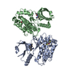
 unidentified baculovirus
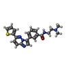
 Mass: 391.489 Da / Num. of mol.: 2 / Source method: obtained synthetically / Formula: C21H21N5OS
 Mass: 154.251 Da / Num. of mol.: 2 / Source method: isolated from a natural source / Formula: C4H10O2S2
 Mass: 18.015 Da / Num. of mol.: 259 / Source method: isolated from a natural source / Formula: H2O
 Sample preparation
 / Beamline: 5.0.2 / Wavelength: 1 Å
 Processing