 Movie
Movie Controller


+ Open data

- Basic information
| Entry | Database: PDB / ID: 3orx | ||||||
|---|---|---|---|---|---|---|---|
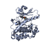
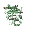
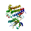
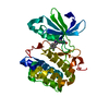
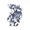
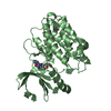
| Title | PDK1 mutant bound to allosteric disulfide fragment inhibitor 1F8 | ||||||
 Components Components | 3-phosphoinositide-dependent protein kinase 1 | ||||||
 Keywords Keywords | TRANSFERASE/TRANSFERASE INHIBITOR / PIF pocket / C-helix / activation loop / AGC kinase / Transferase / Allosteric inhibitor / Phosphorylation / allostery / disulfide / kinase / PDK1 / TRANSFERASE-TRANSFERASE INHIBITOR complex | ||||||
| Function / homology |  Function and homology information Function and homology informationintracellular signaling cassette / 3-phosphoinositide-dependent protein kinase activity / Activation of AKT2 / regulation of mast cell degranulation / type B pancreatic cell development / negative regulation of toll-like receptor signaling pathway / RSK activation / positive regulation of vascular endothelial cell proliferation / regulation of canonical NF-kappaB signal transduction / phospholipase activator activity ...intracellular signaling cassette / 3-phosphoinositide-dependent protein kinase activity / Activation of AKT2 / regulation of mast cell degranulation / type B pancreatic cell development / negative regulation of toll-like receptor signaling pathway / RSK activation / positive regulation of vascular endothelial cell proliferation / regulation of canonical NF-kappaB signal transduction / phospholipase activator activity / positive regulation of sprouting angiogenesis / Constitutive Signaling by AKT1 E17K in Cancer / negative regulation of cardiac muscle cell apoptotic process / Role of LAT2/NTAL/LAB on calcium mobilization / CD28 dependent PI3K/Akt signaling / negative regulation of endothelial cell apoptotic process / positive regulation of blood vessel endothelial cell migration / SARS-CoV-2 targets host intracellular signalling and regulatory pathways / Estrogen-stimulated signaling through PRKCZ / extrinsic apoptotic signaling pathway / vascular endothelial cell response to laminar fluid shear stress / SARS-CoV-1 targets host intracellular signalling and regulatory pathways / RHO GTPases activate PKNs / GPVI-mediated activation cascade / phospholipase binding / T cell costimulation / Integrin signaling / cell projection / insulin-like growth factor receptor signaling pathway / cellular response to epidermal growth factor stimulus / positive regulation of release of sequestered calcium ion into cytosol / VEGFR2 mediated cell proliferation / VEGFR2 mediated vascular permeability / positive regulation of protein localization to plasma membrane / calcium-mediated signaling / phosphatidylinositol 3-kinase/protein kinase B signal transduction / negative regulation of transforming growth factor beta receptor signaling pathway / CLEC7A (Dectin-1) signaling / epidermal growth factor receptor signaling pathway / FCERI mediated NF-kB activation / cellular response to insulin stimulus / positive regulation of angiogenesis / insulin receptor signaling pathway / Regulation of TP53 Degradation / PIP3 activates AKT signaling / protein autophosphorylation / Downstream TCR signaling / G beta:gamma signalling through PI3Kgamma / cell migration / actin cytoskeleton organization / cytoplasmic vesicle / High laminar flow shear stress activates signaling by PIEZO1 and PECAM1:CDH5:KDR in endothelial cells / protein phosphorylation / positive regulation of phosphatidylinositol 3-kinase/protein kinase B signal transduction / non-specific serine/threonine protein kinase / postsynaptic density / intracellular signal transduction / protein serine kinase activity / focal adhesion / protein serine/threonine kinase activity / DNA-templated transcription / ATP binding / nucleus / plasma membrane / cytosol / cytoplasm Similarity search - Function | ||||||
| Biological species |  Homo sapiens (human) Homo sapiens (human) | ||||||
| Method |  X-RAY DIFFRACTION / SYNCHROTRON / MOLECULAR REPLACEMENT / Resolution: 2.2044 Å X-RAY DIFFRACTION / SYNCHROTRON / MOLECULAR REPLACEMENT / Resolution: 2.2044 Å | ||||||
 Authors Authors | Sadowsky, J.D. / Wells, J.A. | ||||||
 Citation Citation | Journal: Proc.Natl.Acad.Sci.USA / Year: 2011 Title: Turning a protein kinase on or off from a single allosteric site via disulfide trapping. Authors: Sadowsky, J.D. / Burlingame, M.A. / Wolan, D.W. / McClendon, C.L. / Jacobson, M.P. / Wells, J.A. | ||||||
| History |
|
- Structure visualization
| Structure viewer | Molecule: MolmilJmol/JSmol |
|---|
- Downloads & links
-Download
| PDBx/mmCIF format | 3orx.cif.gz | 480.4 KB | Display | PDBx/mmCIF format |
|---|---|---|---|---|
| PDB format | pdb3orx.ent.gz | 387.8 KB | Display | PDB format |
| PDBx/mmJSON format | 3orx.json.gz | Tree view | PDBx/mmJSON format | |
| Others |  Other downloads Other downloads |
-Validation report
| Arichive directory | https://data.pdbj.org/pub/pdb/validation_reports/or/3orxftp://data.pdbj.org/pub/pdb/validation_reports/or/3orx | HTTPS FTP |
|---|
-Related structure data
| Related structure data |  3orzC 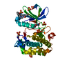 3otuC 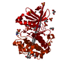 1h1wS S: Starting model for refinement C: citing same article ( |
|---|---|
| Similar structure data |


-Links
 PDBj
PDBj


- Assembly
| Deposited unit | 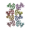
| |||||||||||||||||||||||||||||||||||||||||||||||||||||||||
|---|---|---|---|---|---|---|---|---|---|---|---|---|---|---|---|---|---|---|---|---|---|---|---|---|---|---|---|---|---|---|---|---|---|---|---|---|---|---|---|---|---|---|---|---|---|---|---|---|---|---|---|---|---|---|---|---|---|---|
| 1 | 
| |||||||||||||||||||||||||||||||||||||||||||||||||||||||||
| 2 | 
| |||||||||||||||||||||||||||||||||||||||||||||||||||||||||
| 3 | 
| |||||||||||||||||||||||||||||||||||||||||||||||||||||||||
| 4 | 
| |||||||||||||||||||||||||||||||||||||||||||||||||||||||||
| 5 | 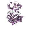
| |||||||||||||||||||||||||||||||||||||||||||||||||||||||||
| 6 | 
| |||||||||||||||||||||||||||||||||||||||||||||||||||||||||
| 7 | 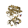
| |||||||||||||||||||||||||||||||||||||||||||||||||||||||||
| 8 | 
| |||||||||||||||||||||||||||||||||||||||||||||||||||||||||
| Unit cell |
| |||||||||||||||||||||||||||||||||||||||||||||||||||||||||
| Noncrystallographic symmetry (NCS) | NCS domain:
NCS domain segments:
NCS ensembles :
|
-Components
| #1: Protein | Mass: 36177.457 Da / Num. of mol.: 8 / Fragment: Catalytic domain (UNP residues 51-359) / Mutation: T148C Source method: isolated from a genetically manipulated source Source: (gene. exp.) Homo sapiens (human) / Gene: PDK1, PDPK1 / Production host:   Spodoptera frugiperda (fall armyworm) / Strain (production host): SF21 Spodoptera frugiperda (fall armyworm) / Strain (production host): SF21References: UniProt: O15530, non-specific serine/threonine protein kinase #2: Chemical | ChemComp-1F8 /   Mass: 235.302 Da / Num. of mol.: 8 / Source method: obtained synthetically / Formula: C12H13NO2S Mass: 235.302 Da / Num. of mol.: 8 / Source method: obtained synthetically / Formula: C12H13NO2S#3: Chemical | ChemComp-CL /   Mass: 35.453 Da / Num. of mol.: 4 / Source method: obtained synthetically / Formula: Cl Mass: 35.453 Da / Num. of mol.: 4 / Source method: obtained synthetically / Formula: Cl#4: Water | ChemComp-HOH / |  Mass: 18.015 Da / Num. of mol.: 1294 / Source method: isolated from a natural source / Formula: H2O Mass: 18.015 Da / Num. of mol.: 1294 / Source method: isolated from a natural source / Formula: H2OHas protein modification | Y | |
|---|
-Experimental details
-Experiment
| Experiment | Method: X-RAY DIFFRACTION / Number of used crystals: 1 |
|---|
- Sample preparation
| Crystal | Density Matthews: 2.34 Å3/Da / Density % sol: 47.41 % |
|---|---|
| Crystal grow | Temperature: 277 K / Method: vapor diffusion, hanging drop / pH: 6.5 Details: 25% PEG 3350, 0.2M lithium sulfate, 0.1M Bis-Tris, pH 6.5, VAPOR DIFFUSION, HANGING DROP, temperature 277K |
-Data collection
| Diffraction | Mean temperature: 100 K |
|---|---|
| Diffraction source | Source: SYNCHROTRON / Site: ALS  / Beamline: 8.3.1 / Wavelength: 1.11589 Å / Beamline: 8.3.1 / Wavelength: 1.11589 Å |
| Detector | Type: ADSC QUANTUM 315r / Detector: CCD / Date: Dec 3, 2009 |
| Radiation | Protocol: SINGLE WAVELENGTH / Monochromatic (M) / Laue (L): M / Scattering type: x-ray |
| Radiation wavelength | Wavelength: 1.11589 Å / Relative weight: 1 |
| Reflection | Resolution: 2.2→50 Å / Num. all: 128125 / Num. obs: 128125 / % possible obs: 97.2 % / Redundancy: 1.8 % / Biso Wilson estimate: 31.1 Å2 / Rmerge(I) obs: 0.067 / Rsym value: 0.067 / Net I/σ(I): 12.765 |
| Reflection shell | Resolution: 2.2→2.28 Å / Redundancy: 1.8 % / Rmerge(I) obs: 0.303 / Mean I/σ(I) obs: 2.761 / Num. unique all: 12662 / Rsym value: 0.303 / % possible all: 96.2 |
- Processing
| Software |
| |||||||||||||||||||||||||||||||||||||||||||||||||||||||||||||||||||||||||||||||||||||||||||||||||||||||||||||||||||||||||||||||||||||||||||||||||||||||||||||||||||||||||||||||||||||||||||||||||||||||||||||||||||||||||
|---|---|---|---|---|---|---|---|---|---|---|---|---|---|---|---|---|---|---|---|---|---|---|---|---|---|---|---|---|---|---|---|---|---|---|---|---|---|---|---|---|---|---|---|---|---|---|---|---|---|---|---|---|---|---|---|---|---|---|---|---|---|---|---|---|---|---|---|---|---|---|---|---|---|---|---|---|---|---|---|---|---|---|---|---|---|---|---|---|---|---|---|---|---|---|---|---|---|---|---|---|---|---|---|---|---|---|---|---|---|---|---|---|---|---|---|---|---|---|---|---|---|---|---|---|---|---|---|---|---|---|---|---|---|---|---|---|---|---|---|---|---|---|---|---|---|---|---|---|---|---|---|---|---|---|---|---|---|---|---|---|---|---|---|---|---|---|---|---|---|---|---|---|---|---|---|---|---|---|---|---|---|---|---|---|---|---|---|---|---|---|---|---|---|---|---|---|---|---|---|---|---|---|---|---|---|---|---|---|---|---|---|---|---|---|---|---|---|---|
| Refinement | Method to determine structure: MOLECULAR REPLACEMENT Starting model: PDB ENTRY 1H1W Resolution: 2.2044→45.247 Å / SU ML: 0.31 / Cross valid method: THROUGHOUT / σ(F): 1.97 / Phase error: 25.12 / Stereochemistry target values: ML
| |||||||||||||||||||||||||||||||||||||||||||||||||||||||||||||||||||||||||||||||||||||||||||||||||||||||||||||||||||||||||||||||||||||||||||||||||||||||||||||||||||||||||||||||||||||||||||||||||||||||||||||||||||||||||
| Solvent computation | Shrinkage radii: 0.9 Å / VDW probe radii: 1.11 Å / Solvent model: FLAT BULK SOLVENT MODEL / Bsol: 50.482 Å2 / ksol: 0.34 e/Å3 | |||||||||||||||||||||||||||||||||||||||||||||||||||||||||||||||||||||||||||||||||||||||||||||||||||||||||||||||||||||||||||||||||||||||||||||||||||||||||||||||||||||||||||||||||||||||||||||||||||||||||||||||||||||||||
| Displacement parameters |
| |||||||||||||||||||||||||||||||||||||||||||||||||||||||||||||||||||||||||||||||||||||||||||||||||||||||||||||||||||||||||||||||||||||||||||||||||||||||||||||||||||||||||||||||||||||||||||||||||||||||||||||||||||||||||
| Refinement step | Cycle: LAST / Resolution: 2.2044→45.247 Å
| |||||||||||||||||||||||||||||||||||||||||||||||||||||||||||||||||||||||||||||||||||||||||||||||||||||||||||||||||||||||||||||||||||||||||||||||||||||||||||||||||||||||||||||||||||||||||||||||||||||||||||||||||||||||||
| Refine LS restraints |
| |||||||||||||||||||||||||||||||||||||||||||||||||||||||||||||||||||||||||||||||||||||||||||||||||||||||||||||||||||||||||||||||||||||||||||||||||||||||||||||||||||||||||||||||||||||||||||||||||||||||||||||||||||||||||
| Refine LS restraints NCS |
| |||||||||||||||||||||||||||||||||||||||||||||||||||||||||||||||||||||||||||||||||||||||||||||||||||||||||||||||||||||||||||||||||||||||||||||||||||||||||||||||||||||||||||||||||||||||||||||||||||||||||||||||||||||||||
| LS refinement shell |
|
