 Movie
Movie Controller

[English] 日本語
 Yorodumi
- PDB-1h1w: High resolution crystal structure of the human PDK1 catalytic domain -
+ Open data

- Basic information
| Entry | Database: PDB / ID: 1h1w | ||||||
|---|---|---|---|---|---|---|---|
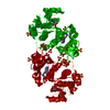
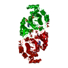
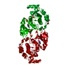
| Title | High resolution crystal structure of the human PDK1 catalytic domain | ||||||
 Components Components | 3-PHOSPHOINOSITIDE DEPENDENT PROTEIN KINASE-1 | ||||||
 Keywords Keywords | TRANSFERASE / PHOSPHOINOSITIDE DEPENDENT PROTEIN KINASE / PKA / AGC KINASE ACTIVATION / PIF-POCKET / PI3-KINASE SIGNALLING / SERINE/THREONINE-PROTEIN KINASE / ATP-BINDING | ||||||
| Function / homology |  Function and homology information Function and homology informationintracellular signaling cassette / 3-phosphoinositide-dependent protein kinase activity / Activation of AKT2 / regulation of mast cell degranulation / type B pancreatic cell development / negative regulation of toll-like receptor signaling pathway / RSK activation / positive regulation of vascular endothelial cell proliferation / regulation of canonical NF-kappaB signal transduction / phospholipase activator activity ...intracellular signaling cassette / 3-phosphoinositide-dependent protein kinase activity / Activation of AKT2 / regulation of mast cell degranulation / type B pancreatic cell development / negative regulation of toll-like receptor signaling pathway / RSK activation / positive regulation of vascular endothelial cell proliferation / regulation of canonical NF-kappaB signal transduction / phospholipase activator activity / positive regulation of sprouting angiogenesis / Constitutive Signaling by AKT1 E17K in Cancer / negative regulation of cardiac muscle cell apoptotic process / Role of LAT2/NTAL/LAB on calcium mobilization / CD28 dependent PI3K/Akt signaling / negative regulation of endothelial cell apoptotic process / positive regulation of blood vessel endothelial cell migration / SARS-CoV-2 targets host intracellular signalling and regulatory pathways / Estrogen-stimulated signaling through PRKCZ / extrinsic apoptotic signaling pathway / vascular endothelial cell response to laminar fluid shear stress / SARS-CoV-1 targets host intracellular signalling and regulatory pathways / RHO GTPases activate PKNs / GPVI-mediated activation cascade / phospholipase binding / T cell costimulation / Integrin signaling / cell projection / insulin-like growth factor receptor signaling pathway / cellular response to epidermal growth factor stimulus / positive regulation of release of sequestered calcium ion into cytosol / VEGFR2 mediated cell proliferation / VEGFR2 mediated vascular permeability / positive regulation of protein localization to plasma membrane / calcium-mediated signaling / phosphatidylinositol 3-kinase/protein kinase B signal transduction / negative regulation of transforming growth factor beta receptor signaling pathway / CLEC7A (Dectin-1) signaling / epidermal growth factor receptor signaling pathway / FCERI mediated NF-kB activation / cellular response to insulin stimulus / positive regulation of angiogenesis / insulin receptor signaling pathway / Regulation of TP53 Degradation / PIP3 activates AKT signaling / protein autophosphorylation / Downstream TCR signaling / G beta:gamma signalling through PI3Kgamma / cell migration / actin cytoskeleton organization / cytoplasmic vesicle / High laminar flow shear stress activates signaling by PIEZO1 and PECAM1:CDH5:KDR in endothelial cells / protein phosphorylation / positive regulation of phosphatidylinositol 3-kinase/protein kinase B signal transduction / non-specific serine/threonine protein kinase / postsynaptic density / intracellular signal transduction / protein serine kinase activity / focal adhesion / protein serine/threonine kinase activity / DNA-templated transcription / ATP binding / nucleus / plasma membrane / cytosol / cytoplasm Similarity search - Function | ||||||
| Biological species |  HOMO SAPIENS (human) HOMO SAPIENS (human) | ||||||
| Method |  X-RAY DIFFRACTION / SYNCHROTRON / MOLECULAR REPLACEMENT / Resolution: 2 Å X-RAY DIFFRACTION / SYNCHROTRON / MOLECULAR REPLACEMENT / Resolution: 2 Å | ||||||
 Authors Authors | Biondi, R.M. / Komander, D. / Thomas, C.C. / Lizcano, J.M. / Deak, M. / Alessi, D.R. / Van Aalten, D.M.F. | ||||||
 Citation Citation | Journal: Embo J. / Year: 2003 Title: High Resolution Crystal Structure of the Human Pdk1 Catalytic Domain Defines the Regulatory Phosphopeptide Docking Site Authors: Biondi, R.M. / Komander, D. / Thomas, C.C. / Lizcano, J.M. / Deak, M. / Alessi, D.R. / Van Aalten, D.M.F. | ||||||
| History |
|
- Structure visualization
| Structure viewer | Molecule: MolmilJmol/JSmol |
|---|
- Downloads & links
-Download
| PDBx/mmCIF format | 1h1w.cif.gz | 80 KB | Display | PDBx/mmCIF format |
|---|---|---|---|---|
| PDB format | pdb1h1w.ent.gz | 59 KB | Display | PDB format |
| PDBx/mmJSON format | 1h1w.json.gz | Tree view | PDBx/mmJSON format | |
| Others |  Other downloads Other downloads |
-Validation report
| Arichive directory | https://data.pdbj.org/pub/pdb/validation_reports/h1/1h1wftp://data.pdbj.org/pub/pdb/validation_reports/h1/1h1w | HTTPS FTP |
|---|
-Related structure data
| Related structure data | 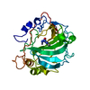 1ydbS S: Starting model for refinement |
|---|---|
| Similar structure data |


-Links
 PDBj
PDBj


- Assembly
| Deposited unit | 
| ||||||||
|---|---|---|---|---|---|---|---|---|---|
| 1 | 
| ||||||||
| Unit cell |
| ||||||||
| Components on special symmetry positions |
|
-Components
| #1: Protein | Mass: 33452.469 Da / Num. of mol.: 1 / Fragment: KINASE CATALYTIC DOMAIN, RESIDUES 71-359 Source method: isolated from a genetically manipulated source Details: ACTIVATION LOOP PHOSPHORYLATION (SER241) / Source: (gene. exp.) HOMO SAPIENS (human) / Production host:   SPODOPTERA FRUGIPERDA (fall armyworm) / Strain (production host): SF21 SPODOPTERA FRUGIPERDA (fall armyworm) / Strain (production host): SF21References: UniProt: O15530, non-specific serine/threonine protein kinase | ||||||||
|---|---|---|---|---|---|---|---|---|---|
| #2: Chemical | ChemComp-GOL /   Mass: 92.094 Da / Num. of mol.: 8 / Source method: obtained synthetically / Formula: C3H8O3 Mass: 92.094 Da / Num. of mol.: 8 / Source method: obtained synthetically / Formula: C3H8O3#3: Chemical | ChemComp-SO4 /   Mass: 96.063 Da / Num. of mol.: 5 / Source method: obtained synthetically / Formula: SO4 Mass: 96.063 Da / Num. of mol.: 5 / Source method: obtained synthetically / Formula: SO4#4: Chemical | ChemComp-ATP / |   Mass: 507.181 Da / Num. of mol.: 1 / Source method: obtained synthetically / Formula: C10H16N5O13P3 / Comment: ATP, energy-carrying molecule*YM Mass: 507.181 Da / Num. of mol.: 1 / Source method: obtained synthetically / Formula: C10H16N5O13P3 / Comment: ATP, energy-carrying molecule*YM#5: Water | ChemComp-HOH / |  Mass: 18.015 Da / Num. of mol.: 200 / Source method: isolated from a natural source / Formula: H2O Mass: 18.015 Da / Num. of mol.: 200 / Source method: isolated from a natural source / Formula: H2OHas protein modification | Y | |
-Experimental details
-Experiment
| Experiment | Method: X-RAY DIFFRACTION / Number of used crystals: 1 |
|---|
- Sample preparation
| Crystal | Density Matthews: 2.81 Å3/Da / Density % sol: 55.9 % | ||||||||||||||||||||||||||||||||||||||||||||||||
|---|---|---|---|---|---|---|---|---|---|---|---|---|---|---|---|---|---|---|---|---|---|---|---|---|---|---|---|---|---|---|---|---|---|---|---|---|---|---|---|---|---|---|---|---|---|---|---|---|---|
| Crystal grow | pH: 8.5 Details: 0.1 M TRIS/HCL PH 8.5, 2.0 M AMMONIUM SULPHATE, 16.6 MM ATP | ||||||||||||||||||||||||||||||||||||||||||||||||
| Crystal grow | *PLUS Temperature: 20 ℃ / pH: 8.5 / Method: vapor diffusion, sitting drop | ||||||||||||||||||||||||||||||||||||||||||||||||
| Components of the solutions | *PLUS
|
-Data collection
| Diffraction | Mean temperature: 100 K |
|---|---|
| Diffraction source | Source: SYNCHROTRON / Site: ESRF  / Beamline: ID14-1 / Wavelength: 0.934 / Beamline: ID14-1 / Wavelength: 0.934 |
| Detector | Type: ADSC CCD / Detector: CCD / Date: Mar 4, 2002 / Details: MIRRORS |
| Radiation | Monochromator: THIN DIAMOND CRYSTAL / Protocol: SINGLE WAVELENGTH / Monochromatic (M) / Laue (L): M / Scattering type: x-ray |
| Radiation wavelength | Wavelength: 0.934 Å / Relative weight: 1 |
| Reflection | Resolution: 2→25 Å / Num. obs: 27643 / % possible obs: 98 % / Redundancy: 2.8 % / Biso Wilson estimate: 12.6 Å2 / Rmerge(I) obs: 0.091 / Net I/σ(I): 7.3 |
| Reflection shell | Resolution: 2→2.07 Å / Redundancy: 2.5 % / Rmerge(I) obs: 0.454 / Mean I/σ(I) obs: 2 / % possible all: 93.5 |
| Reflection | *PLUS Highest resolution: 2 Å / Lowest resolution: 25 Å / Redundancy: 2.8 % / Num. measured all: 77315 / Rmerge(I) obs: 0.091 |
| Reflection shell | *PLUS Highest resolution: 2 Å / % possible obs: 93.5 % / Redundancy: 2.5 % / Rmerge(I) obs: 0.454 / Mean I/σ(I) obs: 2 |
- Processing
| Software |
| ||||||||||||||||||||||||||||||||||||||||||||||||||||||||||||||||||||||||||||||||
|---|---|---|---|---|---|---|---|---|---|---|---|---|---|---|---|---|---|---|---|---|---|---|---|---|---|---|---|---|---|---|---|---|---|---|---|---|---|---|---|---|---|---|---|---|---|---|---|---|---|---|---|---|---|---|---|---|---|---|---|---|---|---|---|---|---|---|---|---|---|---|---|---|---|---|---|---|---|---|---|---|---|
| Refinement | Method to determine structure: MOLECULAR REPLACEMENT Starting model: PDB ENTRY 1YDB Resolution: 2→24.44 Å / Rfactor Rfree error: 0.009 / Data cutoff high absF: 2010867.11 / Data cutoff low absF: 0 / Isotropic thermal model: RESTRAINED / Cross valid method: THROUGHOUT / σ(F): 0
| ||||||||||||||||||||||||||||||||||||||||||||||||||||||||||||||||||||||||||||||||
| Solvent computation | Solvent model: FLAT MODEL / Bsol: 51.6939 Å2 / ksol: 0.380238 e/Å3 | ||||||||||||||||||||||||||||||||||||||||||||||||||||||||||||||||||||||||||||||||
| Displacement parameters | Biso mean: 27.9 Å2
| ||||||||||||||||||||||||||||||||||||||||||||||||||||||||||||||||||||||||||||||||
| Refine analyze |
| ||||||||||||||||||||||||||||||||||||||||||||||||||||||||||||||||||||||||||||||||
| Refinement step | Cycle: LAST / Resolution: 2→24.44 Å
| ||||||||||||||||||||||||||||||||||||||||||||||||||||||||||||||||||||||||||||||||
| Refine LS restraints |
| ||||||||||||||||||||||||||||||||||||||||||||||||||||||||||||||||||||||||||||||||
| LS refinement shell | Resolution: 2→2.13 Å / Rfactor Rfree error: 0.03 / Total num. of bins used: 6
| ||||||||||||||||||||||||||||||||||||||||||||||||||||||||||||||||||||||||||||||||
| Xplor file |
| ||||||||||||||||||||||||||||||||||||||||||||||||||||||||||||||||||||||||||||||||
| Refinement | *PLUS Highest resolution: 2 Å / Lowest resolution: 25 Å / Rfactor Rfree: 0.222 / Rfactor Rwork: 0.195 | ||||||||||||||||||||||||||||||||||||||||||||||||||||||||||||||||||||||||||||||||
| Solvent computation | *PLUS | ||||||||||||||||||||||||||||||||||||||||||||||||||||||||||||||||||||||||||||||||
| Displacement parameters | *PLUS | ||||||||||||||||||||||||||||||||||||||||||||||||||||||||||||||||||||||||||||||||
| Refine LS restraints | *PLUS
|
