 Movie
Movie Controller

[English] 日本語
 Yorodumi
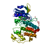
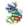
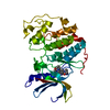
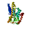
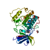
- PDB-4fkg: Crystal structure of the cdk2 in complex with aminopyrazole inhibitor -
+ Open data

- Basic information
| Entry | Database: PDB / ID: 4fkg | ||||||
|---|---|---|---|---|---|---|---|
| Title | Crystal structure of the cdk2 in complex with aminopyrazole inhibitor | ||||||
 Components Components | Cyclin-dependent kinase 2 | ||||||
 Keywords Keywords | TRANSFERASE/TRANSFERASE INHIBITOR / ALPHA AND BETA PROTEIN (A+B) / TRANSFERASE / TRANSFERASE-TRANSFERASE INHIBITOR COMPLEX | ||||||
| Function / homology |  Function and homology information Function and homology informationcyclin A1-CDK2 complex / cyclin E2-CDK2 complex / cyclin E1-CDK2 complex / cyclin A2-CDK2 complex / positive regulation of DNA-templated DNA replication initiation / G2 Phase / Y chromosome / cyclin-dependent protein kinase activity / regulation of heterochromatin organization / Phosphorylation of proteins involved in G1/S transition by active Cyclin E:Cdk2 complexes ...cyclin A1-CDK2 complex / cyclin E2-CDK2 complex / cyclin E1-CDK2 complex / cyclin A2-CDK2 complex / positive regulation of DNA-templated DNA replication initiation / G2 Phase / Y chromosome / cyclin-dependent protein kinase activity / regulation of heterochromatin organization / Phosphorylation of proteins involved in G1/S transition by active Cyclin E:Cdk2 complexes / positive regulation of heterochromatin formation / p53-Dependent G1 DNA Damage Response / X chromosome / PTK6 Regulates Cell Cycle / regulation of anaphase-promoting complex-dependent catabolic process / Defective binding of RB1 mutants to E2F1,(E2F2, E2F3) / centriole replication / Regulation of APC/C activators between G1/S and early anaphase / telomere maintenance in response to DNA damage / centrosome duplication / G0 and Early G1 / Telomere Extension By Telomerase / Activation of the pre-replicative complex / cyclin-dependent kinase / cyclin-dependent protein serine/threonine kinase activity / TP53 Regulates Transcription of Genes Involved in G1 Cell Cycle Arrest / Regulation of MITF-M-dependent genes involved in cell cycle and proliferation / Activation of ATR in response to replication stress / Cajal body / Cyclin E associated events during G1/S transition / Cyclin A:Cdk2-associated events at S phase entry / cyclin-dependent protein kinase holoenzyme complex / Cyclin A/B1/B2 associated events during G2/M transition / regulation of G2/M transition of mitotic cell cycle / condensed chromosome / mitotic G1 DNA damage checkpoint signaling / negative regulation of protein localization to chromatin / cellular response to nitric oxide / post-translational protein modification / regulation of mitotic cell cycle / cyclin binding / positive regulation of DNA replication / peptidyl-serine phosphorylation / male germ cell nucleus / meiotic cell cycle / potassium ion transport / G1/S transition of mitotic cell cycle / DNA Damage/Telomere Stress Induced Senescence / Meiotic recombination / G2/M transition of mitotic cell cycle / CDK-mediated phosphorylation and removal of Cdc6 / Transcriptional regulation of granulopoiesis / SCF(Skp2)-mediated degradation of p27/p21 / Orc1 removal from chromatin / cellular senescence / Cyclin D associated events in G1 / Regulation of TP53 Degradation / nuclear envelope / Factors involved in megakaryocyte development and platelet production / regulation of gene expression / Processing of DNA double-strand break ends / Senescence-Associated Secretory Phenotype (SASP) / transcription regulator complex / Regulation of TP53 Activity through Phosphorylation / protein phosphorylation / Ras protein signal transduction / DNA replication / chromosome, telomeric region / endosome / chromatin remodeling / protein domain specific binding / protein serine kinase activity / cell division / DNA repair / protein serine/threonine kinase activity / positive regulation of cell population proliferation / centrosome / positive regulation of DNA-templated transcription / DNA-templated transcription / magnesium ion binding / negative regulation of transcription by RNA polymerase II / signal transduction / nucleoplasm / ATP binding / nucleus / cytoplasm / cytosol Similarity search - Function | ||||||
| Biological species |  Homo sapiens (human) Homo sapiens (human) | ||||||
| Method |  X-RAY DIFFRACTION / SYNCHROTRON / MOLECULAR REPLACEMENT / Resolution: 1.51 Å X-RAY DIFFRACTION / SYNCHROTRON / MOLECULAR REPLACEMENT / Resolution: 1.51 Å | ||||||
 Authors Authors | Kang, Y.N. / Stuckey, J.A. | ||||||
 Citation Citation | Journal: To be Published Title: Crystal structure of the cdk2 in complex with aminopyrazole inhibitor Authors: Kang, Y.N. / Stuckey, J.A. | ||||||
| History |
|
- Structure visualization
| Structure viewer | Molecule: MolmilJmol/JSmol |
|---|
- Downloads & links
-Download
| PDBx/mmCIF format | 4fkg.cif.gz | 138 KB | Display | PDBx/mmCIF format |
|---|---|---|---|---|
| PDB format | pdb4fkg.ent.gz | 107.6 KB | Display | PDB format |
| PDBx/mmJSON format | 4fkg.json.gz | Tree view | PDBx/mmJSON format | |
| Others |  Other downloads Other downloads |
-Validation report
| Arichive directory | https://data.pdbj.org/pub/pdb/validation_reports/fk/4fkgftp://data.pdbj.org/pub/pdb/validation_reports/fk/4fkg | HTTPS FTP |
|---|
-Related structure data
| Related structure data |  4ek4C  4ek5C  4ek6C  4fkjC 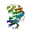 2r3iS C: citing same article ( S: Starting model for refinement |
|---|---|
| Similar structure data |


-Links
 PDBj
PDBj


- Assembly
| Deposited unit | 
| ||||||||
|---|---|---|---|---|---|---|---|---|---|
| 1 |
| ||||||||
| Unit cell |
|
-Components
| #1: Protein | Mass: 34002.527 Da / Num. of mol.: 1 Source method: isolated from a genetically manipulated source Source: (gene. exp.) Homo sapiens (human) / Gene: CDK2, CDKN2 / Plasmid: PFB1 / Cell line (production host): HIGH-FIVE / Production host:  TRICHOPLUSIA NI (cabbage looper) / References: UniProt: P24941, cyclin-dependent kinase TRICHOPLUSIA NI (cabbage looper) / References: UniProt: P24941, cyclin-dependent kinase |
|---|---|
| #2: Chemical | ChemComp-4CK /   Mass: 271.271 Da / Num. of mol.: 1 / Source method: obtained synthetically / Formula: C14H13N3O3 Mass: 271.271 Da / Num. of mol.: 1 / Source method: obtained synthetically / Formula: C14H13N3O3 |
| #3: Water | ChemComp-HOH /  Mass: 18.015 Da / Num. of mol.: 214 / Source method: isolated from a natural source / Formula: H2O Mass: 18.015 Da / Num. of mol.: 214 / Source method: isolated from a natural source / Formula: H2O |
| Has protein modification | Y |
-Experimental details
-Experiment
| Experiment | Method: X-RAY DIFFRACTION / Number of used crystals: 1 |
|---|
- Sample preparation
| Crystal | Density Matthews: 2.03 Å3/Da / Density % sol: 39.49 % |
|---|---|
| Crystal grow | Temperature: 293.15 K / Method: vapor diffusion, sitting drop / pH: 7.8 Details: 15-20% PEG3350, 0.2M AMMONIUM ACETATE, 0.1M HEPES, VAPOR DIFFUSION, SITTING DROP, TEMPERATURE 293.15K, VAPOR DIFFUSION, SITTING DROP, pH 7.8 |
-Data collection
| Diffraction | Mean temperature: 100 K |
|---|---|
| Diffraction source | Source: SYNCHROTRON / Site: APS  / Beamline: 21-ID-G / Wavelength: 0.97856 / Wavelength: 0.97856 Å / Beamline: 21-ID-G / Wavelength: 0.97856 / Wavelength: 0.97856 Å |
| Detector | Type: MARMOSAIC 300 mm CCD / Detector: CCD / Date: Oct 17, 2009 / Details: MIRRORS |
| Radiation | Monochromator: DIAMOND [111] / Protocol: SINGLE WAVELENGTH / Monochromatic (M) / Laue (L): M / Scattering type: x-ray |
| Radiation wavelength | Wavelength: 0.97856 Å / Relative weight: 1 |
| Reflection | Resolution: 1.51→50 Å / Num. all: 44045 / Num. obs: 43429 / % possible obs: 98.6 % / Observed criterion σ(I): -3 / Redundancy: 6.9 % / Rmerge(I) obs: 0.043 / Net I/σ(I): 15.4 |
| Reflection shell | Resolution: 1.51→1.54 Å / Redundancy: 3.7 % / Rmerge(I) obs: 0.397 / % possible all: 78.6 |
- Processing
| Software |
| |||||||||||||||||||||||||||||||||||||||||||||||||||||||||||||||||||||||||||||||||||||||||||||||||||||||||||||||||||||||||||||||||||||||||||||||||||||||||||||||||||||||||||||||
|---|---|---|---|---|---|---|---|---|---|---|---|---|---|---|---|---|---|---|---|---|---|---|---|---|---|---|---|---|---|---|---|---|---|---|---|---|---|---|---|---|---|---|---|---|---|---|---|---|---|---|---|---|---|---|---|---|---|---|---|---|---|---|---|---|---|---|---|---|---|---|---|---|---|---|---|---|---|---|---|---|---|---|---|---|---|---|---|---|---|---|---|---|---|---|---|---|---|---|---|---|---|---|---|---|---|---|---|---|---|---|---|---|---|---|---|---|---|---|---|---|---|---|---|---|---|---|---|---|---|---|---|---|---|---|---|---|---|---|---|---|---|---|---|---|---|---|---|---|---|---|---|---|---|---|---|---|---|---|---|---|---|---|---|---|---|---|---|---|---|---|---|---|---|---|---|---|
| Refinement | Method to determine structure: MOLECULAR REPLACEMENT Starting model: PDB ENTRY 2R3I Resolution: 1.51→36.82 Å / Cor.coef. Fo:Fc: 0.961 / Cor.coef. Fo:Fc free: 0.949 / SU R Cruickshank DPI: 0.08 / Cross valid method: THROUGHOUT / σ(F): 0 / Stereochemistry target values: MAXIMUM LIKELIHOOD
| |||||||||||||||||||||||||||||||||||||||||||||||||||||||||||||||||||||||||||||||||||||||||||||||||||||||||||||||||||||||||||||||||||||||||||||||||||||||||||||||||||||||||||||||
| Displacement parameters | Biso mean: 29.16 Å2
| |||||||||||||||||||||||||||||||||||||||||||||||||||||||||||||||||||||||||||||||||||||||||||||||||||||||||||||||||||||||||||||||||||||||||||||||||||||||||||||||||||||||||||||||
| Refine analyze | Luzzati coordinate error obs: 0.19 Å | |||||||||||||||||||||||||||||||||||||||||||||||||||||||||||||||||||||||||||||||||||||||||||||||||||||||||||||||||||||||||||||||||||||||||||||||||||||||||||||||||||||||||||||||
| Refinement step | Cycle: LAST / Resolution: 1.51→36.82 Å
| |||||||||||||||||||||||||||||||||||||||||||||||||||||||||||||||||||||||||||||||||||||||||||||||||||||||||||||||||||||||||||||||||||||||||||||||||||||||||||||||||||||||||||||||
| Refine LS restraints |
| |||||||||||||||||||||||||||||||||||||||||||||||||||||||||||||||||||||||||||||||||||||||||||||||||||||||||||||||||||||||||||||||||||||||||||||||||||||||||||||||||||||||||||||||
| LS refinement shell | Resolution: 1.51→1.55 Å / Total num. of bins used: 20
| |||||||||||||||||||||||||||||||||||||||||||||||||||||||||||||||||||||||||||||||||||||||||||||||||||||||||||||||||||||||||||||||||||||||||||||||||||||||||||||||||||||||||||||||
| Refinement TLS params. | Method: refined / Refine-ID: X-RAY DIFFRACTION
| |||||||||||||||||||||||||||||||||||||||||||||||||||||||||||||||||||||||||||||||||||||||||||||||||||||||||||||||||||||||||||||||||||||||||||||||||||||||||||||||||||||||||||||||
| Refinement TLS group |
|
