 Movie
Movie Controller

[English] 日本語
 Yorodumi
- PDB-3sfc: Structure-Based Optimization of Potent 4- and 6-Azaindole-3-Carbo... -
+ Open data

- Basic information
| Entry | Database: PDB / ID: 3sfc | ||||||
|---|---|---|---|---|---|---|---|
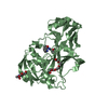
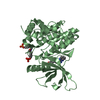
| Title | Structure-Based Optimization of Potent 4- and 6-Azaindole-3-Carboxamides as Renin Inhibitors | ||||||
 Components Components | Renin | ||||||
 Keywords Keywords | HYDROLASE/HYDROLASE INHIBITOR / RENIN HUMAN / ASPARTYL PROTEASE / RENIN INHIBITION / HYPERTENSION / beta barrel / pepsin-like protease / glycosylation / extracellular space / HYDROLASE-HYDROLASE INHIBITOR complex | ||||||
| Function / homology |  Function and homology information Function and homology informationrenin / mesonephros development / juxtaglomerular apparatus development / renin-angiotensin regulation of aldosterone production / response to cGMP / drinking behavior / response to immobilization stress / regulation of MAPK cascade / amyloid-beta metabolic process / response to cAMP ...renin / mesonephros development / juxtaglomerular apparatus development / renin-angiotensin regulation of aldosterone production / response to cGMP / drinking behavior / response to immobilization stress / regulation of MAPK cascade / amyloid-beta metabolic process / response to cAMP / Metabolism of Angiotensinogen to Angiotensins / angiotensin maturation / insulin-like growth factor receptor binding / cell maturation / hormone-mediated signaling pathway / kidney development / male gonad development / regulation of blood pressure / cellular response to xenobiotic stimulus / apical part of cell / peptidase activity / response to lipopolysaccharide / aspartic-type endopeptidase activity / signaling receptor binding / proteolysis / : / extracellular region / plasma membrane Similarity search - Function | ||||||
| Biological species |  Homo sapiens (human) Homo sapiens (human) | ||||||
| Method |  X-RAY DIFFRACTION / SYNCHROTRON / FOURIER SYNTHESIS / Resolution: 2.1 Å X-RAY DIFFRACTION / SYNCHROTRON / FOURIER SYNTHESIS / Resolution: 2.1 Å | ||||||
 Authors Authors | Scheiper, B. / Matter, H. / Steinhagen, H. / Bocskei, Z. / Fleury, V. / McCort, G. | ||||||
 Citation Citation | Journal: Bioorg.Med.Chem.Lett. / Year: 2011 Title: Structure-based optimization of potent 4- and 6-azaindole-3-carboxamides as renin inhibitors. Authors: Scheiper, B. / Matter, H. / Steinhagen, H. / Bocskei, Z. / Fleury, V. / McCort, G. #1: Journal: Bioorg.Med.Chem.Lett. / Year: 2010Title: Discovery and optimization of a new class of potent and non-chiral indole-3-carboxamide-based renin inhibitors. Authors: Scheiper, B. / Matter, H. / Steinhagen, H. / Stilz, U. / Bocskei, Z. / Fleury, V. / McCort, G. | ||||||
| History |
|
- Structure visualization
| Structure viewer | Molecule: MolmilJmol/JSmol |
|---|
- Downloads & links
-Download
| PDBx/mmCIF format | 3sfc.cif.gz | 289.2 KB | Display | PDBx/mmCIF format |
|---|---|---|---|---|
| PDB format | pdb3sfc.ent.gz | 235.7 KB | Display | PDB format |
| PDBx/mmJSON format | 3sfc.json.gz | Tree view | PDBx/mmJSON format | |
| Others |  Other downloads Other downloads |
-Validation report
| Arichive directory | https://data.pdbj.org/pub/pdb/validation_reports/sf/3sfcftp://data.pdbj.org/pub/pdb/validation_reports/sf/3sfc | HTTPS FTP |
|---|
-Related structure data
| Related structure data | |
|---|---|
| Similar structure data |


-Links
 PDBj
PDBj


- Assembly
| Deposited unit | 
| ||||||||
|---|---|---|---|---|---|---|---|---|---|
| 1 | 
| ||||||||
| 2 | 
| ||||||||
| 3 | 
| ||||||||
| 4 | 
| ||||||||
| 5 | 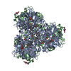
| ||||||||
| Unit cell |
|
-Components
| #1: Protein | Mass: 37267.008 Da / Num. of mol.: 2 / Fragment: unp residues 67-406 Source method: isolated from a genetically manipulated source Source: (gene. exp.) Homo sapiens (human) / Gene: REN / Cell line (production host): HEK / Production host: HOMO SAPIENS (human) / References: UniProt: P00797, renin#2: Sugar |   Type: D-saccharide, beta linking / Mass: 221.208 Da / Num. of mol.: 2 Type: D-saccharide, beta linking / Mass: 221.208 Da / Num. of mol.: 2Source method: isolated from a genetically manipulated source Formula: C8H15NO6 #3: Chemical | ChemComp-GOL /   Mass: 92.094 Da / Num. of mol.: 4 / Source method: obtained synthetically / Formula: C3H8O3 Mass: 92.094 Da / Num. of mol.: 4 / Source method: obtained synthetically / Formula: C3H8O3#4: Chemical |   Mass: 520.597 Da / Num. of mol.: 3 / Source method: obtained synthetically / Formula: C32H29FN4O2 Mass: 520.597 Da / Num. of mol.: 3 / Source method: obtained synthetically / Formula: C32H29FN4O2#5: Water | ChemComp-HOH / |  Mass: 18.015 Da / Num. of mol.: 483 / Source method: isolated from a natural source / Formula: H2O Mass: 18.015 Da / Num. of mol.: 483 / Source method: isolated from a natural source / Formula: H2OHas protein modification | Y | |
|---|
-Experimental details
-Experiment
| Experiment | Method: X-RAY DIFFRACTION / Number of used crystals: 1 |
|---|
- Sample preparation
| Crystal | Density Matthews: 2.99 Å3/Da / Density % sol: 58.85 % |
|---|---|
| Crystal grow | Temperature: 298 K / Method: vapor diffusion, hanging drop / pH: 4.5 Details: 0.05M CITRATE, 10-12% PEG3350, 0.6M NACL, 20 MG/ML RENIN, VAPOUR DIFFUSION, HANGING DROP, VAPOR DIFFUSION, HANGING DROP, temperature 298K, pH 4.5 |
-Data collection
| Diffraction | Mean temperature: 100 K |
|---|---|
| Diffraction source | Source: SYNCHROTRON / Site: ESRF  / Beamline: ID23-1 / Wavelength: 1.07225 Å / Beamline: ID23-1 / Wavelength: 1.07225 Å |
| Detector | Type: ADSC QUANTUM 315r / Detector: CCD / Date: Dec 6, 2007 |
| Radiation | Monochromator: mirror / Protocol: SINGLE WAVELENGTH / Monochromatic (M) / Laue (L): M / Scattering type: x-ray |
| Radiation wavelength | Wavelength: 1.07225 Å / Relative weight: 1 |
| Reflection | Resolution: 2.1→69.34 Å / Num. all: 51036 / Num. obs: 51036 / % possible obs: 98.2 % / Observed criterion σ(F): 0 / Observed criterion σ(I): -5 / Redundancy: 2.5 % / Biso Wilson estimate: 33.835 Å2 / Rmerge(I) obs: 0.159 / Net I/σ(I): 3.3 |
| Reflection shell | Resolution: 2.1→2.21 Å / Redundancy: 2.6 % / Rmerge(I) obs: 0.779 / Mean I/σ(I) obs: 1 / Rsym value: 0.779 / % possible all: 99.5 |
- Processing
| Software |
| |||||||||||||||||||||||||||||||||||||||||||||||||||||||||||||||||||||||||||||||||||||||||||||||||||||||||||||||||||||||||||||||||||||||||||||||||||||||||||||||||||||||||||||||||||||||||||||||||||||||||||||||||||||||||||||||||
|---|---|---|---|---|---|---|---|---|---|---|---|---|---|---|---|---|---|---|---|---|---|---|---|---|---|---|---|---|---|---|---|---|---|---|---|---|---|---|---|---|---|---|---|---|---|---|---|---|---|---|---|---|---|---|---|---|---|---|---|---|---|---|---|---|---|---|---|---|---|---|---|---|---|---|---|---|---|---|---|---|---|---|---|---|---|---|---|---|---|---|---|---|---|---|---|---|---|---|---|---|---|---|---|---|---|---|---|---|---|---|---|---|---|---|---|---|---|---|---|---|---|---|---|---|---|---|---|---|---|---|---|---|---|---|---|---|---|---|---|---|---|---|---|---|---|---|---|---|---|---|---|---|---|---|---|---|---|---|---|---|---|---|---|---|---|---|---|---|---|---|---|---|---|---|---|---|---|---|---|---|---|---|---|---|---|---|---|---|---|---|---|---|---|---|---|---|---|---|---|---|---|---|---|---|---|---|---|---|---|---|---|---|---|---|---|---|---|---|---|---|---|---|---|---|---|---|
| Refinement | Method to determine structure: FOURIER SYNTHESIS / Resolution: 2.1→62.07 Å / Cor.coef. Fo:Fc: 0.9126 / Cor.coef. Fo:Fc free: 0.9046 / Occupancy max: 1 / Occupancy min: 0.5 / SU R Cruickshank DPI: 0.207 / Cross valid method: THROUGHOUT / σ(F): 0
| |||||||||||||||||||||||||||||||||||||||||||||||||||||||||||||||||||||||||||||||||||||||||||||||||||||||||||||||||||||||||||||||||||||||||||||||||||||||||||||||||||||||||||||||||||||||||||||||||||||||||||||||||||||||||||||||||
| Displacement parameters | Biso max: 188.23 Å2 / Biso mean: 48.9062 Å2 / Biso min: 19.63 Å2
| |||||||||||||||||||||||||||||||||||||||||||||||||||||||||||||||||||||||||||||||||||||||||||||||||||||||||||||||||||||||||||||||||||||||||||||||||||||||||||||||||||||||||||||||||||||||||||||||||||||||||||||||||||||||||||||||||
| Refine analyze | Luzzati coordinate error obs: 0.301 Å | |||||||||||||||||||||||||||||||||||||||||||||||||||||||||||||||||||||||||||||||||||||||||||||||||||||||||||||||||||||||||||||||||||||||||||||||||||||||||||||||||||||||||||||||||||||||||||||||||||||||||||||||||||||||||||||||||
| Refinement step | Cycle: LAST / Resolution: 2.1→62.07 Å
| |||||||||||||||||||||||||||||||||||||||||||||||||||||||||||||||||||||||||||||||||||||||||||||||||||||||||||||||||||||||||||||||||||||||||||||||||||||||||||||||||||||||||||||||||||||||||||||||||||||||||||||||||||||||||||||||||
| Refine LS restraints |
| |||||||||||||||||||||||||||||||||||||||||||||||||||||||||||||||||||||||||||||||||||||||||||||||||||||||||||||||||||||||||||||||||||||||||||||||||||||||||||||||||||||||||||||||||||||||||||||||||||||||||||||||||||||||||||||||||
| LS refinement shell | Resolution: 2.1→2.15 Å / Total num. of bins used: 20
| |||||||||||||||||||||||||||||||||||||||||||||||||||||||||||||||||||||||||||||||||||||||||||||||||||||||||||||||||||||||||||||||||||||||||||||||||||||||||||||||||||||||||||||||||||||||||||||||||||||||||||||||||||||||||||||||||
| Refinement TLS params. | Method: refined / Refine-ID: X-RAY DIFFRACTION
| |||||||||||||||||||||||||||||||||||||||||||||||||||||||||||||||||||||||||||||||||||||||||||||||||||||||||||||||||||||||||||||||||||||||||||||||||||||||||||||||||||||||||||||||||||||||||||||||||||||||||||||||||||||||||||||||||
| Refinement TLS group |
|
