 Movie
Movie Controller

[English] 日本語
 Yorodumi
- PDB-1ls5: Crystal structure of plasmepsin IV from P. falciparum in complex ... -
+ Open data

- Basic information
| Entry | Database: PDB / ID: 1ls5 | |||||||||
|---|---|---|---|---|---|---|---|---|---|---|
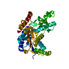
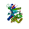
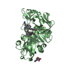
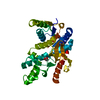
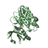
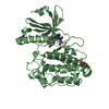
| Title | Crystal structure of plasmepsin IV from P. falciparum in complex with pepstatin A | |||||||||
 Components Components |
| |||||||||
 Keywords Keywords | HYDROLASE/HYDROLASE INHIBITOR / EUKARYOTIC ASPARTIC PROTEINASE / HYDROLASE-HYDROLASE INHIBITOR complex | |||||||||
| Function / homology |  Function and homology information Function and homology informationMHC class II antigen presentation / hemoglobin catabolic process / plasmepsin II / acquisition of nutrients from host / vacuolar lumen / food vacuole / aspartic-type endopeptidase activity / lysosome / proteolysis / membrane Similarity search - Function | |||||||||
| Biological species |   Streptomyces argenteolus subsp. toyonakensis (bacteria) Streptomyces argenteolus subsp. toyonakensis (bacteria) | |||||||||
| Method |  X-RAY DIFFRACTION / MOLECULAR REPLACEMENT / Resolution: 2.8 Å X-RAY DIFFRACTION / MOLECULAR REPLACEMENT / Resolution: 2.8 Å | |||||||||
 Authors Authors | Asojo, O.A. / Gulnik, S.V. / Afonina, E. / Yu, B. / Ellman, J.A. / Haque, T.S. / Silva, A.M. | |||||||||
 Citation Citation | Journal: J.Mol.Biol. / Year: 2003 Title: Novel uncomplexed and complexed structures of plasmepsin II, an aspartic protease from Plasmodium falciparum. Authors: Asojo, O.A. / Gulnik, S.V. / Afonina, E. / Yu, B. / Ellman, J.A. / Haque, T.S. / Silva, A.M. | |||||||||
| History |
|
- Structure visualization
| Structure viewer | Molecule: MolmilJmol/JSmol |
|---|
- Downloads & links
-Download
| PDBx/mmCIF format | 1ls5.cif.gz | 129.8 KB | Display | PDBx/mmCIF format |
|---|---|---|---|---|
| PDB format | pdb1ls5.ent.gz | 104.4 KB | Display | PDB format |
| PDBx/mmJSON format | 1ls5.json.gz | Tree view | PDBx/mmJSON format | |
| Others |  Other downloads Other downloads |
-Validation report
| Arichive directory | https://data.pdbj.org/pub/pdb/validation_reports/ls/1ls5ftp://data.pdbj.org/pub/pdb/validation_reports/ls/1ls5 | HTTPS FTP |
|---|
-Related structure data
| Related structure data |  1lf3C  1lf4C  1smeS S: Starting model for refinement C: citing same article ( |
|---|---|
| Similar structure data |


-Links
 PDBj
PDBj
- Assembly
| Deposited unit | 
| ||||||||
|---|---|---|---|---|---|---|---|---|---|
| 1 | 
| ||||||||
| 2 | 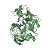
| ||||||||
| 3 | 
| ||||||||
| 4 |
| ||||||||
| Unit cell |
|
-Components
| #1: Protein | Mass: 36985.457 Da / Num. of mol.: 2 Source method: isolated from a genetically manipulated source Source: (gene. exp.) Production host: #2: Protein/peptide | 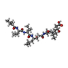  Type: Oligopeptide / Class: Enzyme inhibitor / Mass: 685.891 Da / Num. of mol.: 2 / Source method: obtained synthetically Type: Oligopeptide / Class: Enzyme inhibitor / Mass: 685.891 Da / Num. of mol.: 2 / Source method: obtained syntheticallySource: (synth.) Streptomyces argenteolus subsp. toyonakensis (bacteria)References: Pepstatin #3: Water | ChemComp-HOH / |  Mass: 18.015 Da / Num. of mol.: 28 / Source method: isolated from a natural source / Formula: H2O Mass: 18.015 Da / Num. of mol.: 28 / Source method: isolated from a natural source / Formula: H2OHas protein modification | Y | |
|---|
-Experimental details
-Experiment
| Experiment | Method: X-RAY DIFFRACTION / Number of used crystals: 1 |
|---|
- Sample preparation
| Crystal | Density Matthews: 2.5 Å3/Da / Density % sol: 50.81 % |
|---|---|
| Crystal grow | pH: 5.5 / Details: pH 5.50 |
-Data collection
| Diffraction | Mean temperature: 90 K |
|---|---|
| Diffraction source | Source: ROTATING ANODE / Type: RIGAKU RU200 / Wavelength: 1.5418 / Wavelength: 1.5418 Å |
| Detector | Type: MAR scanner 300 mm plate / Detector: IMAGE PLATE / Date: Mar 1, 2000 |
| Radiation | Monochromator: DOUBLE FOCUSING MIRROR SYSTEM / Protocol: SINGLE WAVELENGTH / Monochromatic (M) / Laue (L): M / Scattering type: x-ray |
| Radiation wavelength | Wavelength: 1.5418 Å / Relative weight: 1 |
| Reflection | Resolution: 2.8→20 Å / Num. obs: 15253 / % possible obs: 79 % / Observed criterion σ(I): 1 / Rmerge(I) obs: 0.096 / Rsym value: 0.083 / Net I/σ(I): 8.5 |
- Processing
| Software |
| ||||||||||||||||||||||||||||||||||||||||||||||||||||||||||||
|---|---|---|---|---|---|---|---|---|---|---|---|---|---|---|---|---|---|---|---|---|---|---|---|---|---|---|---|---|---|---|---|---|---|---|---|---|---|---|---|---|---|---|---|---|---|---|---|---|---|---|---|---|---|---|---|---|---|---|---|---|---|
| Refinement | Method to determine structure: MOLECULAR REPLACEMENT Starting model: PLASMEPSIN II, PDB ENTRY 1SME Resolution: 2.8→10 Å / σ(F): 3
| ||||||||||||||||||||||||||||||||||||||||||||||||||||||||||||
| Refinement step | Cycle: LAST / Resolution: 2.8→10 Å
| ||||||||||||||||||||||||||||||||||||||||||||||||||||||||||||
| Refine LS restraints |
|
