 Movie
Movie Controller

[English] 日本語
 Yorodumi
- PDB-1fak: HUMAN TISSUE FACTOR COMPLEXED WITH COAGULATION FACTOR VIIA INHIBI... -
+ Open data

- Basic information
| Entry | Database: PDB / ID: 1fak | ||||||
|---|---|---|---|---|---|---|---|
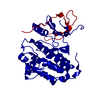
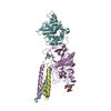
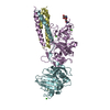
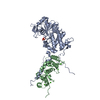
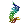
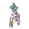
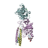
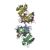
| Title | HUMAN TISSUE FACTOR COMPLEXED WITH COAGULATION FACTOR VIIA INHIBITED WITH A BPTI-MUTANT | ||||||
 Components Components |
| ||||||
 Keywords Keywords | BLOOD CLOTTING / COMPLEX(SERINE PROTEASE-COFACTOR-LIGAND) / BLOOD COAGULATION / SERINE PROTEASE / COMPLEX / CO-FACTOR / RECEPTOR ENZYME / INHIBITOR / GLA / EGF / COMPLEX (SERINE PROTEASE-COFACTOR-LIGAND) | ||||||
| Function / homology |  Function and homology information Function and homology informationactivation of plasma proteins involved in acute inflammatory response / activation of blood coagulation via clotting cascade / coagulation factor VIIa / response to Thyroid stimulating hormone / response to astaxanthin / response to thyrotropin-releasing hormone / response to 2,3,7,8-tetrachlorodibenzodioxine / response to carbon dioxide / serine-type peptidase complex / response to genistein ...activation of plasma proteins involved in acute inflammatory response / activation of blood coagulation via clotting cascade / coagulation factor VIIa / response to Thyroid stimulating hormone / response to astaxanthin / response to thyrotropin-releasing hormone / response to 2,3,7,8-tetrachlorodibenzodioxine / response to carbon dioxide / serine-type peptidase complex / response to genistein / sulfate binding / response to vitamin K / positive regulation of platelet-derived growth factor receptor signaling pathway / positive regulation of leukocyte chemotaxis / negative regulation of platelet aggregation / response to thyroxine / NGF-stimulated transcription / zymogen binding / response to cholesterol / potassium channel inhibitor activity / cytokine receptor activity / positive regulation of positive chemotaxis / : / response to growth hormone / molecular function inhibitor activity / negative regulation of thrombin-activated receptor signaling pathway / positive regulation of blood coagulation / animal organ regeneration / positive regulation of endothelial cell apoptotic process / positive regulation of TOR signaling / Transport of gamma-carboxylated protein precursors from the endoplasmic reticulum to the Golgi apparatus / Gamma-carboxylation of protein precursors / Removal of aminoterminal propeptides from gamma-carboxylated proteins / serine protease inhibitor complex / positive regulation of endothelial cell proliferation / BMAL1:CLOCK,NPAS2 activates circadian expression / serine-type peptidase activity / positive regulation of interleukin-8 production / serine-type endopeptidase inhibitor activity / circadian rhythm / protein processing / phospholipid binding / response to estrogen / Golgi lumen / cytokine-mediated signaling pathway / positive regulation of angiogenesis / blood coagulation / response to estradiol / extracellular matrix / protease binding / vesicle / response to hypoxia / positive regulation of cell migration / endoplasmic reticulum lumen / signaling receptor binding / serine-type endopeptidase activity / external side of plasma membrane / calcium ion binding / positive regulation of gene expression / cell surface / : / extracellular region / membrane / plasma membrane Similarity search - Function | ||||||
| Biological species |  Homo sapiens (human) Homo sapiens (human) | ||||||
| Method |  X-RAY DIFFRACTION / SYNCHROTRON / MOLECULAR REPLACEMENT / Resolution: 2.1 Å X-RAY DIFFRACTION / SYNCHROTRON / MOLECULAR REPLACEMENT / Resolution: 2.1 Å | ||||||
 Authors Authors | Zhang, E. / St Charles, R. / Tulinsky, A. | ||||||
 Citation Citation | Journal: J.Mol.Biol. / Year: 1999 Title: Structure of extracellular tissue factor complexed with factor VIIa inhibited with a BPTI mutant. Authors: Zhang, E. / St Charles, R. / Tulinsky, A. | ||||||
| History |
|
- Structure visualization
| Structure viewer | Molecule: MolmilJmol/JSmol |
|---|
- Downloads & links
-Download
| PDBx/mmCIF format | 1fak.cif.gz | 139.4 KB | Display | PDBx/mmCIF format |
|---|---|---|---|---|
| PDB format | pdb1fak.ent.gz | 108.3 KB | Display | PDB format |
| PDBx/mmJSON format | 1fak.json.gz | Tree view | PDBx/mmJSON format | |
| Others |  Other downloads Other downloads |
-Validation report
| Arichive directory | https://data.pdbj.org/pub/pdb/validation_reports/fa/1fakftp://data.pdbj.org/pub/pdb/validation_reports/fa/1fak | HTTPS FTP |
|---|
-Related structure data
| Similar structure data |
|---|


-Links
 PDBj
PDBj


- Assembly
| Deposited unit | 
| ||||||||
|---|---|---|---|---|---|---|---|---|---|
| 1 |
| ||||||||
| Unit cell |
|
-Components
-PROTEIN (BLOOD COAGULATION FACTOR ... , 2 types, 2 molecules LH
| #1: Protein | Mass: 17487.076 Da / Num. of mol.: 1 / Fragment: LIGHT CHAIN Source method: isolated from a genetically manipulated source Source: (gene. exp.) Homo sapiens (human) / Cell line (production host): KIDNEY CELLS (BHK) / Production host:   Cricetulus griseus (Chinese hamster) / References: UniProt: P08709, coagulation factor VIIa Cricetulus griseus (Chinese hamster) / References: UniProt: P08709, coagulation factor VIIa |
|---|---|
| #2: Protein | Mass: 28103.256 Da / Num. of mol.: 1 / Fragment: HEAVY CHAIN Source method: isolated from a genetically manipulated source Source: (gene. exp.) Homo sapiens (human) / Cell line (production host): KIDNEY CELLS (BHK) / Production host: Cricetulus griseus (Chinese hamster) / References: UniProt: P08709, coagulation factor VIIa |
-Protein , 2 types, 2 molecules TI
| #3: Protein | Mass: 23417.053 Da / Num. of mol.: 1 Source method: isolated from a genetically manipulated source Source: (gene. exp.) Homo sapiens (human) / Production host:  |
|---|---|
| #4: Protein | Mass: 6299.163 Da / Num. of mol.: 1 / Source method: obtained synthetically / Details: Chemically synthesized / References: UniProt: P00974 |
-Sugars , 2 types, 2 molecules 

| #5: Sugar | ChemComp-GLC /  Type: D-saccharide, alpha linking / Mass: 180.156 Da / Num. of mol.: 1 Type: D-saccharide, alpha linking / Mass: 180.156 Da / Num. of mol.: 1Source method: isolated from a genetically manipulated source Formula: C6H12O6 |
|---|---|
| #6: Sugar | ChemComp-FUC /  Type: L-saccharide, alpha linking / Mass: 164.156 Da / Num. of mol.: 1 Type: L-saccharide, alpha linking / Mass: 164.156 Da / Num. of mol.: 1Source method: isolated from a genetically manipulated source Formula: C6H12O5 |
-Non-polymers , 2 types, 342 molecules 

| #7: Chemical |  Mass: 40.078 Da / Num. of mol.: 2 / Source method: obtained synthetically / Formula: Ca Mass: 40.078 Da / Num. of mol.: 2 / Source method: obtained synthetically / Formula: Ca#8: Water | ChemComp-HOH / | Mass: 18.015 Da / Num. of mol.: 340 / Source method: isolated from a natural source / Formula: H2O |
|---|
-Experimental details
-Experiment
| Experiment | Method: X-RAY DIFFRACTION / Number of used crystals: 1 |
|---|
- Sample preparation
| Crystal | Density Matthews: 3.68 Å3/Da / Density % sol: 64 % | |||||||||||||||||||||||||||||||||||||||||||||||||||||||||||||||||||||||||||||
|---|---|---|---|---|---|---|---|---|---|---|---|---|---|---|---|---|---|---|---|---|---|---|---|---|---|---|---|---|---|---|---|---|---|---|---|---|---|---|---|---|---|---|---|---|---|---|---|---|---|---|---|---|---|---|---|---|---|---|---|---|---|---|---|---|---|---|---|---|---|---|---|---|---|---|---|---|---|---|
| Crystal grow | pH: 5.6 / Details: pH 5.6 | |||||||||||||||||||||||||||||||||||||||||||||||||||||||||||||||||||||||||||||
| Crystal grow | *PLUS Method: vapor diffusion, hanging drop | |||||||||||||||||||||||||||||||||||||||||||||||||||||||||||||||||||||||||||||
| Components of the solutions | *PLUS
|
-Data collection
| Diffraction | Mean temperature: 100 K |
|---|---|
| Diffraction source | Source: SYNCHROTRON / Site: APS  / Beamline: 17-ID / Wavelength: 0.89 / Beamline: 17-ID / Wavelength: 0.89 |
| Detector | Type: SIEMENS / Detector: CCD |
| Radiation | Protocol: SINGLE WAVELENGTH / Monochromatic (M) / Laue (L): M / Scattering type: x-ray |
| Radiation wavelength | Wavelength: 0.89 Å / Relative weight: 1 |
| Reflection | Resolution: 2.1→20 Å / Num. obs: 61209 / % possible obs: 87 % / Observed criterion σ(I): 1 / Redundancy: 2.35 % / Rmerge(I) obs: 0.094 |
| Reflection | *PLUS Num. obs: 70671 / Num. measured all: 166318 |
| Reflection shell | *PLUS Highest resolution: 2.1 Å / Lowest resolution: 2.2 Å / % possible obs: 78 % / Num. unique obs: 61209 |
- Processing
| Software |
| ||||||||||||||||||||||||||||||||||||||||||||||||||||||||||||||||||||||||||||||||||||
|---|---|---|---|---|---|---|---|---|---|---|---|---|---|---|---|---|---|---|---|---|---|---|---|---|---|---|---|---|---|---|---|---|---|---|---|---|---|---|---|---|---|---|---|---|---|---|---|---|---|---|---|---|---|---|---|---|---|---|---|---|---|---|---|---|---|---|---|---|---|---|---|---|---|---|---|---|---|---|---|---|---|---|---|---|---|
| Refinement | Method to determine structure: MOLECULAR REPLACEMENT / Resolution: 2.1→9 Å / σ(F): 0 /
| ||||||||||||||||||||||||||||||||||||||||||||||||||||||||||||||||||||||||||||||||||||
| Refinement step | Cycle: LAST / Resolution: 2.1→9 Å
| ||||||||||||||||||||||||||||||||||||||||||||||||||||||||||||||||||||||||||||||||||||
| Refine LS restraints |
| ||||||||||||||||||||||||||||||||||||||||||||||||||||||||||||||||||||||||||||||||||||
| Software | *PLUS Name: PROLSQ / Classification: refinement | ||||||||||||||||||||||||||||||||||||||||||||||||||||||||||||||||||||||||||||||||||||
| Refinement | *PLUS Highest resolution: 2.1 Å / σ(F): 0 | ||||||||||||||||||||||||||||||||||||||||||||||||||||||||||||||||||||||||||||||||||||
| Solvent computation | *PLUS | ||||||||||||||||||||||||||||||||||||||||||||||||||||||||||||||||||||||||||||||||||||
| Displacement parameters | *PLUS | ||||||||||||||||||||||||||||||||||||||||||||||||||||||||||||||||||||||||||||||||||||
| Refine LS restraints | *PLUS
|
