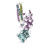
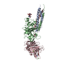
[English] 日本語
Yorodumi
- PDB-2hlo: Crystal Structure of Fragment D-dimer from Human Fibrin Complexed... -

+
Open data


ID or keywords:

Loading...

-
Basic information

Entry
Database: PDB / ID: 2hlo
TitleCrystal Structure of Fragment D-dimer from Human Fibrin Complexed with Gly-hydroxyPro-Arg-Pro-amide
Components
  • Fibrinogen alpha chain
  • Fibrinogen beta chain
  • GLY-HYP-ARG-PRO-AMIDE PEPTIDE LIGAND
  • Isoform Gamma-A of Fibrinogen gamma chain
KeywordsBLOOD CLOTTING / Coiled-coil / knob-hole interactions
Function / homology
Function and homology information


platelet maturation / blood coagulation, common pathway / induction of bacterial agglutination / fibrinogen complex / Regulation of TLR by endogenous ligand / platelet alpha granule / blood coagulation, fibrin clot formation / cellular response to leptin stimulus / cellular response to interleukin-6 / MyD88 deficiency (TLR2/4) ...platelet maturation / blood coagulation, common pathway / induction of bacterial agglutination / fibrinogen complex / Regulation of TLR by endogenous ligand / platelet alpha granule / blood coagulation, fibrin clot formation / cellular response to leptin stimulus / cellular response to interleukin-6 / MyD88 deficiency (TLR2/4) / positive regulation of heterotypic cell-cell adhesion / IRAK4 deficiency (TLR2/4) / MyD88:MAL(TIRAP) cascade initiated on plasma membrane / extracellular matrix structural constituent / plasminogen activation / p130Cas linkage to MAPK signaling for integrins / positive regulation of peptide hormone secretion / positive regulation of exocytosis / GRB2:SOS provides linkage to MAPK signaling for Integrins / protein secretion / protein polymerization / cellular response to interleukin-1 / Integrin cell surface interactions / negative regulation of endothelial cell apoptotic process / Common Pathway of Fibrin Clot Formation / negative regulation of extrinsic apoptotic signaling pathway via death domain receptors / positive regulation of substrate adhesion-dependent cell spreading / positive regulation of vasoconstriction / cell adhesion molecule binding / fibrinolysis / Integrin signaling / cell-matrix adhesion / platelet alpha granule lumen / positive regulation of protein secretion / Post-translational protein phosphorylation / Signaling by high-kinase activity BRAF mutants / MAP2K and MAPK activation / platelet aggregation / response to calcium ion / Signaling by RAF1 mutants / Signaling by moderate kinase activity BRAF mutants / Paradoxical activation of RAF signaling by kinase inactive BRAF / Signaling downstream of RAS mutants / Regulation of Insulin-like Growth Factor (IGF) transport and uptake by Insulin-like Growth Factor Binding Proteins (IGFBPs) / Signaling by BRAF and RAF1 fusions / extracellular vesicle / Platelet degranulation / cell cortex / ER-Phagosome pathway / protein-folding chaperone binding / protein-containing complex assembly / collagen-containing extracellular matrix / adaptive immune response / positive regulation of ERK1 and ERK2 cascade / blood microparticle / Amyloid fiber formation / endoplasmic reticulum lumen / external side of plasma membrane / innate immune response / signaling receptor binding / synapse / structural molecule activity / cell surface / endoplasmic reticulum / extracellular space / extracellular exosome / extracellular region / identical protein binding / metal ion binding / plasma membrane
Similarity search - Function
Single alpha-helices involved in coiled-coils or other helix-helix interfaces - #50 / Fibrinogen alpha C domain / Fibrinogen alpha C domain / Fibrinogen, alpha/beta/gamma chain, coiled coil domain / Fibrinogen alpha chain / Fibrinogen alpha/beta chain family / Fibrinogen alpha/beta chain family / Gamma-fibrinogen Carboxyl Terminal Fragment; domain 2 / Gamma-fibrinogen Carboxyl Terminal Fragment, domain 2 / Gamma Fibrinogen; Chain A, domain 1 ...Single alpha-helices involved in coiled-coils or other helix-helix interfaces - #50 / Fibrinogen alpha C domain / Fibrinogen alpha C domain / Fibrinogen, alpha/beta/gamma chain, coiled coil domain / Fibrinogen alpha chain / Fibrinogen alpha/beta chain family / Fibrinogen alpha/beta chain family / Gamma-fibrinogen Carboxyl Terminal Fragment; domain 2 / Gamma-fibrinogen Carboxyl Terminal Fragment, domain 2 / Gamma Fibrinogen; Chain A, domain 1 / Gamma Fibrinogen, chain A, domain 1 / Fibrinogen, conserved site / Fibrinogen C-terminal domain signature. / Fibrinogen-related domains (FReDs) / Fibrinogen, alpha/beta/gamma chain, C-terminal globular, subdomain 1 / Fibrinogen beta and gamma chains, C-terminal globular domain / Fibrinogen, alpha/beta/gamma chain, C-terminal globular domain / Fibrinogen-like, C-terminal / Fibrinogen C-terminal domain profile. / Single alpha-helices involved in coiled-coils or other helix-helix interfaces / Few Secondary Structures / Irregular / Alpha-Beta Complex / Up-down Bundle / Mainly Alpha / Alpha Beta
Similarity search - Domain/homology
Fibrinogen alpha chain / Fibrinogen beta chain / Fibrinogen gamma chain
Similarity search - Component
Biological speciesHomo sapiens (human)
MethodX-RAY DIFFRACTION / SYNCHROTRON / MOLECULAR REPLACEMENT / Resolution: 2.6 Å
AuthorsDoolittle, R.F. / Kollman, J.M. / Chen, A. / Pandi, L.
History
DepositionJul 8, 2006Deposition site: RCSB / Processing site: RCSB
Revision 1.0Jul 17, 2007Provider: repository / Type: Initial release
Revision 1.1May 1, 2008Group: Version format compliance
Revision 1.2Jul 13, 2011Group: Non-polymer description / Version format compliance
Revision 2.0Jul 29, 2020Group: Atomic model / Data collection ...Atomic model / Data collection / Derived calculations / Structure summary
Category: atom_site / chem_comp ...atom_site / chem_comp / entity / pdbx_branch_scheme / pdbx_chem_comp_identifier / pdbx_entity_branch / pdbx_entity_branch_descriptor / pdbx_entity_branch_link / pdbx_entity_branch_list / pdbx_entity_nonpoly / pdbx_nonpoly_scheme / pdbx_struct_assembly_gen / pdbx_struct_conn_angle / struct_asym / struct_conn / struct_site / struct_site_gen
Item: _atom_site.B_iso_or_equiv / _atom_site.Cartn_x ..._atom_site.B_iso_or_equiv / _atom_site.Cartn_x / _atom_site.Cartn_y / _atom_site.Cartn_z / _atom_site.auth_asym_id / _atom_site.auth_atom_id / _atom_site.auth_comp_id / _atom_site.auth_seq_id / _atom_site.label_asym_id / _atom_site.label_atom_id / _atom_site.label_comp_id / _atom_site.label_entity_id / _atom_site.type_symbol / _chem_comp.name / _chem_comp.type / _entity.formula_weight / _entity.pdbx_description / _entity.pdbx_number_of_molecules / _entity.src_method / _entity.type / _pdbx_struct_assembly_gen.asym_id_list / _pdbx_struct_conn_angle.ptnr1_auth_asym_id / _pdbx_struct_conn_angle.ptnr1_auth_comp_id / _pdbx_struct_conn_angle.ptnr1_auth_seq_id / _pdbx_struct_conn_angle.ptnr1_label_asym_id / _pdbx_struct_conn_angle.ptnr1_label_atom_id / _pdbx_struct_conn_angle.ptnr1_label_comp_id / _pdbx_struct_conn_angle.ptnr1_label_seq_id / _pdbx_struct_conn_angle.ptnr2_auth_seq_id / _pdbx_struct_conn_angle.ptnr2_label_asym_id / _pdbx_struct_conn_angle.ptnr3_auth_asym_id / _pdbx_struct_conn_angle.ptnr3_auth_comp_id / _pdbx_struct_conn_angle.ptnr3_auth_seq_id / _pdbx_struct_conn_angle.ptnr3_label_asym_id / _pdbx_struct_conn_angle.ptnr3_label_atom_id / _pdbx_struct_conn_angle.ptnr3_label_comp_id / _pdbx_struct_conn_angle.ptnr3_label_seq_id / _pdbx_struct_conn_angle.value / _struct_conn.conn_type_id / _struct_conn.id / _struct_conn.pdbx_dist_value / _struct_conn.pdbx_leaving_atom_flag / _struct_conn.pdbx_role / _struct_conn.ptnr1_auth_asym_id / _struct_conn.ptnr1_auth_comp_id / _struct_conn.ptnr1_auth_seq_id / _struct_conn.ptnr1_label_asym_id / _struct_conn.ptnr1_label_atom_id / _struct_conn.ptnr1_label_comp_id / _struct_conn.ptnr1_label_seq_id / _struct_conn.ptnr2_auth_asym_id / _struct_conn.ptnr2_auth_comp_id / _struct_conn.ptnr2_auth_seq_id / _struct_conn.ptnr2_label_asym_id / _struct_conn.ptnr2_label_atom_id / _struct_conn.ptnr2_label_comp_id / _struct_conn.ptnr2_label_seq_id
Description: Carbohydrate remediation / Provider: repository / Type: Remediation
Revision 2.1Aug 30, 2023Group: Data collection / Database references ...Data collection / Database references / Refinement description / Source and taxonomy / Structure summary
Category: chem_comp / chem_comp_atom ...chem_comp / chem_comp_atom / chem_comp_bond / database_2 / entity / entity_src_nat / pdbx_entity_src_syn / pdbx_initial_refinement_model / struct_ref / struct_ref_seq
Item: _chem_comp.pdbx_synonyms / _database_2.pdbx_DOI ..._chem_comp.pdbx_synonyms / _database_2.pdbx_DOI / _database_2.pdbx_database_accession / _entity.pdbx_description / _entity_src_nat.pdbx_beg_seq_num / _entity_src_nat.pdbx_end_seq_num / _pdbx_entity_src_syn.pdbx_beg_seq_num / _pdbx_entity_src_syn.pdbx_end_seq_num

-
Structure visualization

Structure viewerMolecule:
MolmilJmol/JSmol

Downloads & links

-
Assembly

Deposited unit
A: Fibrinogen alpha chain
B: Fibrinogen beta chain
C: Isoform Gamma-A of Fibrinogen gamma chain
D: Fibrinogen alpha chain
E: Fibrinogen beta chain
F: Isoform Gamma-A of Fibrinogen gamma chain
G: GLY-HYP-ARG-PRO-AMIDE PEPTIDE LIGAND
H: GLY-HYP-ARG-PRO-AMIDE PEPTIDE LIGAND
hetero molecules


Theoretical massNumber of molelcules
Total (without water)171,27217
Polymers170,1428
Non-polymers1,1299
Water3,639202
1
A: Fibrinogen alpha chain
B: Fibrinogen beta chain
C: Isoform Gamma-A of Fibrinogen gamma chain
G: GLY-HYP-ARG-PRO-AMIDE PEPTIDE LIGAND
hetero molecules


Theoretical massNumber of molelcules
Total (without water)85,6569
Polymers85,0714
Non-polymers5855
Water724
TypeNameSymmetry operationNumber
identity operation1_555x,y,z1
2
D: Fibrinogen alpha chain
E: Fibrinogen beta chain
F: Isoform Gamma-A of Fibrinogen gamma chain
H: GLY-HYP-ARG-PRO-AMIDE PEPTIDE LIGAND
hetero molecules


Theoretical massNumber of molelcules
Total (without water)85,6168
Polymers85,0714
Non-polymers5454
Water724
TypeNameSymmetry operationNumber
identity operation1_555x,y,z1
Unit cell
Length a, b, c (Å)54.690, 149.480, 232.930
Angle α, β, γ (deg.)90.00, 90.00, 90.00
Int Tables number19
Space group name H-MP212121

-
Components

-
Protein , 3 types, 6 molecules ADBECF

#1: Protein Fibrinogen alpha chain


Mass: 10244.963 Da / Num. of mol.: 2 / Source method: isolated from a natural source / Source: (natural) Homo sapiens (human) / References: UniProt: P02671
#2: Protein Fibrinogen beta chain


Mass: 37691.992 Da / Num. of mol.: 2 / Source method: isolated from a natural source / Source: (natural) Homo sapiens (human) / References: UniProt: P02675
#3: Protein Isoform Gamma-A of Fibrinogen gamma chain


Mass: 36693.754 Da / Num. of mol.: 2 / Source method: isolated from a natural source / Source: (natural) Homo sapiens (human) / References: UniProt: P02679

-
Protein/peptide , 1 types, 2 molecules GH

#4: Protein/peptide GLY-HYP-ARG-PRO-AMIDE PEPTIDE LIGAND


Mass: 440.498 Da / Num. of mol.: 2 / Source method: obtained synthetically / Details: The peptide was chemically synthesized.

-
Sugars , 2 types, 2 molecules

#5: Polysaccharide 2-acetamido-2-deoxy-beta-D-glucopyranose-(1-4)-2-acetamido-2-deoxy-beta-D-glucopyranose


Type: oligosaccharide / Mass: 424.401 Da / Num. of mol.: 1
Source method: isolated from a genetically manipulated source
DescriptorTypeProgram
DGlcpNAcb1-4DGlcpNAcb1-Glycam Condensed SequenceGMML 1.0
WURCS=2.0/1,2,1/[a2122h-1b_1-5_2*NCC/3=O]/1-1/a4-b1WURCSPDB2Glycan 1.1.0
[]{[(4+1)][b-D-GlcpNAc]{[(4+1)][b-D-GlcpNAc]{}}}LINUCSPDB-CARE
#6: Polysaccharide 2-acetamido-2-deoxy-alpha-D-glucopyranose-(1-4)-2-acetamido-2-deoxy-beta-D-glucopyranose


Type: oligosaccharide / Mass: 424.401 Da / Num. of mol.: 1
Source method: isolated from a genetically manipulated source
DescriptorTypeProgram
DGlcpNAca1-4DGlcpNAcb1-Glycam Condensed SequenceGMML 1.0
WURCS=2.0/2,2,1/[a2122h-1b_1-5_2*NCC/3=O][a2122h-1a_1-5_2*NCC/3=O]/1-2/a4-b1WURCSPDB2Glycan 1.1.0
[]{[(4+1)][b-D-GlcpNAc]{[(4+1)][a-D-GlcpNAc]{}}}LINUCSPDB-CARE

-
Non-polymers , 2 types, 209 molecules

#7: Chemical
ChemComp-CA / CALCIUM ION


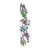
Mass: 40.078 Da / Num. of mol.: 7 / Source method: obtained synthetically / Formula: Ca
#8: Water ChemComp-HOH / water


Mass: 18.015 Da / Num. of mol.: 202 / Source method: isolated from a natural source / Formula: H2O

-
Experimental details

-
Experiment

ExperimentMethod: X-RAY DIFFRACTION / Number of used crystals: 1

-
Sample preparation

CrystalDensity Matthews: 2.8 Å3/Da / Density % sol: 56.03 %
Crystal growTemperature: 295 K / Method: vapor diffusion, sitting drop / pH: 8
Details: 10mg/ml fibrinogen, 2mM Gly-hydroxyPro-Arg-Pro-amide, 7.5mM CaCl2, 10% PEG, 50mM Tris, 2mM sodium azide, pH 8.0, VAPOR DIFFUSION, SITTING DROP, temperature 295K

-
Data collection

DiffractionMean temperature: 100 K
Diffraction sourceSource: SYNCHROTRON / Site: ALS / Beamline: 8.2.1 / Wavelength: 1 Å
DetectorType: ADSC QUANTUM 210 / Detector: CCD / Date: Mar 4, 2005 / Details: KOHZU: Double Crystal Si(111)
RadiationMonochromator: Double crystal, Si(111) / Protocol: SINGLE WAVELENGTH / Monochromatic (M) / Laue (L): M / Scattering type: x-ray
Radiation wavelengthWavelength: 1 Å / Relative weight: 1
ReflectionResolution: 2.6→50 Å / Num. all: 56193 / Num. obs: 56193 / % possible obs: 93.9 % / Observed criterion σ(F): 0 / Observed criterion σ(I): 0 / Redundancy: 6.3 % / Rsym value: 0.128

-
Processing

Software
NameVersionClassification
ADSCQuantumdata collection
AMoREphasing
CNSrefinement
HKL-2000data reduction
SCALEPACKdata scaling
RefinementMethod to determine structure: MOLECULAR REPLACEMENT
Starting model: 1FZF
Resolution: 2.6→30 Å / Cross valid method: CNS / σ(F): 2 / Stereochemistry target values: Engh & Huber
RfactorNum. reflection% reflectionSelection details
Rfree0.2675 2825 -RANDOM
Rwork0.2104 ---
all0.2048 56193 --
obs0.2024 56193 95 %-
Refinement stepCycle: LAST / Resolution: 2.6→30 Å
ProteinNucleic acidLigandSolventTotal
Num. atoms10485 0 63 202 10750
Refine LS restraints
Refine-IDTypeDev ideal
X-RAY DIFFRACTIONc_bond_d0.006177
X-RAY DIFFRACTIONc_bond_d_na
X-RAY DIFFRACTIONc_bond_d_prot
X-RAY DIFFRACTIONc_angle_d1.24396
X-RAY DIFFRACTIONc_angle_d_na
X-RAY DIFFRACTIONc_angle_d_prot
X-RAY DIFFRACTIONc_angle_deg
X-RAY DIFFRACTIONc_angle_deg_na
X-RAY DIFFRACTIONc_angle_deg_prot
X-RAY DIFFRACTIONc_dihedral_angle_d
X-RAY DIFFRACTIONc_dihedral_angle_d_na
X-RAY DIFFRACTIONc_dihedral_angle_d_prot
X-RAY DIFFRACTIONc_improper_angle_d
X-RAY DIFFRACTIONc_improper_angle_d_na
X-RAY DIFFRACTIONc_improper_angle_d_prot
X-RAY DIFFRACTIONc_mcbond_it
X-RAY DIFFRACTIONc_mcangle_it
X-RAY DIFFRACTIONc_scbond_it
X-RAY DIFFRACTIONc_scangle_it
LS refinement shellHighest resolution: 2.6 Å
RfactorNum. reflection% reflection
Rfree0.2675 2825 -
Rwork0.2104 --
obs--93.9 %

+
About Yorodumi

-
News

-
Feb 9, 2022. New format data for meta-information of EMDB entries

New format data for meta-information of EMDB entries

  • Version 3 of the EMDB header file is now the official format.
  • The previous official version 1.9 will be removed from the archive.

Related info.:EMDB header

External links:wwPDB to switch to version 3 of the EMDB data model

-
Aug 12, 2020. Covid-19 info

Covid-19 info

URL: https://pdbjlvh1.pdbj.org/emnavi/covid19.php

New page: Covid-19 featured information page in EM Navigator.

Related info.:Covid-19 info / Mar 5, 2020. Novel coronavirus structure data

+
Mar 5, 2020. Novel coronavirus structure data

Novel coronavirus structure data

Related info.:Yorodumi Speices / Aug 12, 2020. Covid-19 info

External links:COVID-19 featured content - PDBj / Molecule of the Month (242):Coronavirus Proteases

+
Jan 31, 2019. EMDB accession codes are about to change! (news from PDBe EMDB page)

EMDB accession codes are about to change! (news from PDBe EMDB page)

  • The allocation of 4 digits for EMDB accession codes will soon come to an end. Whilst these codes will remain in use, new EMDB accession codes will include an additional digit and will expand incrementally as the available range of codes is exhausted. The current 4-digit format prefixed with “EMD-” (i.e. EMD-XXXX) will advance to a 5-digit format (i.e. EMD-XXXXX), and so on. It is currently estimated that the 4-digit codes will be depleted around Spring 2019, at which point the 5-digit format will come into force.
  • The EM Navigator/Yorodumi systems omit the EMD- prefix.

Related info.:Q: What is EMD? / ID/Accession-code notation in Yorodumi/EM Navigator

External links:EMDB Accession Codes are Changing Soon! / Contact to PDBj

+
Jul 12, 2017. Major update of PDB

Major update of PDB

  • wwPDB released updated PDB data conforming to the new PDBx/mmCIF dictionary.
  • This is a major update changing the version number from 4 to 5, and with Remediation, in which all the entries are updated.
  • In this update, many items about electron microscopy experimental information are reorganized (e.g. em_software).
  • Now, EM Navigator and Yorodumi are based on the updated data.

External links:wwPDB Remediation / Enriched Model Files Conforming to OneDep Data Standards Now Available in the PDB FTP Archive

-
Yorodumi

Thousand views of thousand structures

  • Yorodumi is a browser for structure data from EMDB, PDB, SASBDB, etc.
  • This page is also the successor to EM Navigator detail page, and also detail information page/front-end page for Omokage search.
  • The word "yorodu" (or yorozu) is an old Japanese word meaning "ten thousand". "mi" (miru) is to see.

Related info.:EMDB / PDB / SASBDB / Comparison of 3 databanks / Yorodumi Search / Aug 31, 2016. New EM Navigator & Yorodumi / Yorodumi Papers / Jmol/JSmol / Function and homology information / Changes in new EM Navigator and Yorodumi

Read more