 ムービー
ムービー コントローラー


+ データを開く

- 基本情報
基本情報
| 登録情報 | データベース: PDB / ID: 1jbu | ||||||
|---|---|---|---|---|---|---|---|
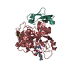
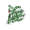
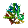
| タイトル | Coagulation Factor VII Zymogen (EGF2/Protease) in Complex with Inhibitory Exosite Peptide A-183 | ||||||
 要素 要素 |
| ||||||
 キーワード キーワード | HYDROLASE/HYDROLASE INHIBITOR / shifted registration / beta-strands / HYDROLASE-HYDROLASE INHIBITOR complex | ||||||
| 機能・相同性 |  機能・相同性情報 機能・相同性情報coagulation factor VIIa / response to Thyroid stimulating hormone / response to astaxanthin / response to thyrotropin-releasing hormone / response to 2,3,7,8-tetrachlorodibenzodioxine / response to carbon dioxide / serine-type peptidase complex / response to genistein / response to vitamin K / positive regulation of platelet-derived growth factor receptor signaling pathway ...coagulation factor VIIa / response to Thyroid stimulating hormone / response to astaxanthin / response to thyrotropin-releasing hormone / response to 2,3,7,8-tetrachlorodibenzodioxine / response to carbon dioxide / serine-type peptidase complex / response to genistein / response to vitamin K / positive regulation of platelet-derived growth factor receptor signaling pathway / positive regulation of leukocyte chemotaxis / response to thyroxine / response to cholesterol / positive regulation of positive chemotaxis / : / response to growth hormone / positive regulation of blood coagulation / animal organ regeneration / positive regulation of TOR signaling / Transport of gamma-carboxylated protein precursors from the endoplasmic reticulum to the Golgi apparatus / Gamma-carboxylation of protein precursors / Removal of aminoterminal propeptides from gamma-carboxylated proteins / BMAL1:CLOCK,NPAS2 activates circadian expression / serine-type peptidase activity / circadian rhythm / protein processing / response to estrogen / Golgi lumen / blood coagulation / response to estradiol / extracellular matrix / vesicle / response to hypoxia / positive regulation of cell migration / endoplasmic reticulum lumen / signaling receptor binding / serine-type endopeptidase activity / calcium ion binding / : / extracellular region / plasma membrane 類似検索 - 分子機能 | ||||||
| 生物種 |  Homo sapiens (ヒト) Homo sapiens (ヒト) | ||||||
| 手法 |  X線回折 / シンクロトロン / 分子置換 / 解像度: 2 Å X線回折 / シンクロトロン / 分子置換 / 解像度: 2 Å | ||||||
 データ登録者 データ登録者 | Eigenbrot, C. / Kirchhofer, D. / Dennis, M.S. / Santell, L. / Lazarus, R.A. / Stamos, J. / Ultsch, M.H. | ||||||
 引用 引用 | ジャーナル: Structure / 年: 2001 タイトル: The factor VII zymogen structure reveals reregistration of beta strands during activation. 著者: Eigenbrot, C. / Kirchhofer, D. / Dennis, M.S. / Santell, L. / Lazarus, R.A. / Stamos, J. / Ultsch, M.H. #1: ジャーナル: To be Publishedタイトル: A Novel Exosite on Coagulation Factor VIIa and its Molecular Interactions with a New Class of Peptide Inhibitors 著者: Roberge, M. / Santell, L. / Dennis, M.S. / Eigenbrot, C. / Dwyer, M.A. / Lazarus, R.A. #2: ジャーナル: Nature / 年: 1996タイトル: The Crystal Structure of the Complex of Blood Coagulation Factor VIIa with Soluble Tissue Factor 著者: Banner, D.W. / D'Arcy, A. / Chene, C. / Winkler, F.K. / Guha, A. / Konigsberg, W.H. / Nemerson, Y. / Kirchhofer, D. #3: ジャーナル: J.Mol.Biol. / 年: 1999タイトル: Structure of Extracellular Tissue Factor Complexed with Factor VIIa Inhibited with a BPTI Mutant 著者: Zhang, E. / St.Charles, R. / Tulinsky, A. #4: ジャーナル: J.STRUCT.BIOL. / 年: 1999タイトル: Crystal Structure of Active Site-inhibited Human Coagulation Factor VIIa (des-Gla) 著者: Kemball-Cook, G. / Johnson, D.J. / Tuddenham, E.G. / Harlos, K. #5: ジャーナル: Proc.Natl.Acad.Sci.USA / 年: 1999タイトル: Structure of Human Factor VIIa and its Implications for the Triggering of Blood Coagulation 著者: Pike, A.C. / Brzozowski, A.M. / Roberts, S.M. / Olsen, O.H. / Persson, E. #6: ジャーナル: Nature / 年: 2000タイトル: Peptide Exosite Inhibitors of Factor VIIa as Anticoagulants 著者: Dennis, M.S. / Eigenbrot, C. / Skelton, N.J. / Ultsch, M.H. / Santell, L. / Dwyer, M.A. / O'Connell, M.P. / Lazarus, R.A. | ||||||
| 履歴 |
| ||||||
| Remark 600 | HETEROGEN The authors are uncertain of the true identity of the het group BEN, and used benzamidine ...HETEROGEN The authors are uncertain of the true identity of the het group BEN, and used benzamidine as a convenience. |
- 構造の表示
構造の表示
| 構造ビューア | 分子: MolmilJmol/JSmol |
|---|
- ダウンロードとリンク
-ダウンロード
| PDBx/mmCIF形式 | 1jbu.cif.gz | 83.2 KB | 表示 | PDBx/mmCIF形式 |
|---|---|---|---|---|
| PDB形式 | pdb1jbu.ent.gz | 60.9 KB | 表示 | PDB形式 |
| PDBx/mmJSON形式 | 1jbu.json.gz | ツリー表示 | PDBx/mmJSON形式 | |
| その他 |  その他のダウンロード その他のダウンロード |
-検証レポート
| アーカイブディレクトリ | https://data.pdbj.org/pub/pdb/validation_reports/jb/1jbuftp://data.pdbj.org/pub/pdb/validation_reports/jb/1jbu | HTTPS FTP |
|---|
-関連構造データ
| 関連構造データ |  1danS S: 精密化の開始モデル |
|---|---|
| 類似構造データ |


-リンク
 PDBj
PDBj


- 集合体
集合体
| 登録構造単位 | 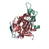
| ||||||||
|---|---|---|---|---|---|---|---|---|---|
| 1 |
| ||||||||
| 2 | 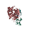
| ||||||||
| 単位格子 |
|
-要素
-COAGULATION FACTOR ... , 2種, 2分子 HL
| #1: タンパク質 | 分子量: 28103.256 Da / 分子数: 1 / 断片: Heavy chain / 由来タイプ: 組換発現 / 由来: (組換発現) Homo sapiens (ヒト) / プラスミド: pAcGP67 / 発現宿主:  Trichoplusia ni (イラクサキンウワバ) / 株 (発現宿主): High-Five / 参照: UniProt: P08709, coagulation factor VIIa Trichoplusia ni (イラクサキンウワバ) / 株 (発現宿主): High-Five / 参照: UniProt: P08709, coagulation factor VIIa |
|---|---|
| #2: タンパク質 | 分子量: 6871.753 Da / 分子数: 1 / 断片: Light chain / 由来タイプ: 組換発現 / 由来: (組換発現) Homo sapiens (ヒト) / プラスミド: pAcGP67 / 発現宿主: Trichoplusia ni (イラクサキンウワバ) / 株 (発現宿主): High-Five / 参照: UniProt: P08709, coagulation factor VIIa |
-タンパク質・ペプチド , 1種, 1分子 X
| #3: タンパク質・ペプチド | 分子量: 2000.189 Da / 分子数: 1 / 由来タイプ: 組換発現 / 由来: (組換発現) |
|---|
-非ポリマー , 3種, 200分子 


| #4: 化合物 | ChemComp-SO4 /  分子量: 96.063 Da / 分子数: 1 / 由来タイプ: 合成 / 式: SO4 分子量: 96.063 Da / 分子数: 1 / 由来タイプ: 合成 / 式: SO4 |
|---|---|
| #5: 化合物 | ChemComp-BEN /  分子量: 120.152 Da / 分子数: 1 / 由来タイプ: 合成 / 式: C7H8N2 分子量: 120.152 Da / 分子数: 1 / 由来タイプ: 合成 / 式: C7H8N2 |
| #6: 水 | ChemComp-HOH / 分子量: 18.015 Da / 分子数: 198 / 由来タイプ: 天然 / 式: H2O |
-詳細
| Has protein modification | Y |
|---|
-実験情報
-実験
| 実験 | 手法: X線回折 / 使用した結晶の数: 1 |
|---|
- 試料調製
試料調製
| 結晶 | マシュー密度: 3.27 Å3/Da / 溶媒含有率: 62.36 % | ||||||||||||||||||||||||||||||||||||||||||||||||||||||
|---|---|---|---|---|---|---|---|---|---|---|---|---|---|---|---|---|---|---|---|---|---|---|---|---|---|---|---|---|---|---|---|---|---|---|---|---|---|---|---|---|---|---|---|---|---|---|---|---|---|---|---|---|---|---|---|
| 結晶化 | 温度: 293 K / 手法: 蒸気拡散法, ハンギングドロップ法 / pH: 7.5 詳細: HEPES, ammonium sulfate, glycerol, PEG 400, benzamidine, calcium chloride, pH 7.5, VAPOR DIFFUSION, HANGING DROP, temperature 293K | ||||||||||||||||||||||||||||||||||||||||||||||||||||||
| 結晶化 | *PLUS | ||||||||||||||||||||||||||||||||||||||||||||||||||||||
| 溶液の組成 | *PLUS
|
-データ収集
| 回折 | 平均測定温度: 100 K |
|---|---|
| 放射光源 | 由来: シンクロトロン / サイト: ALS  / ビームライン: 5.0.2 / 波長: 1.05 Å / ビームライン: 5.0.2 / 波長: 1.05 Å |
| 検出器 | タイプ: ADSC QUANTUM 4 / 検出器: CCD / 日付: 1999年12月8日 |
| 放射 | モノクロメーター: double crystal / プロトコル: SINGLE WAVELENGTH / 単色(M)・ラウエ(L): M / 散乱光タイプ: x-ray |
| 放射波長 | 波長: 1.05 Å / 相対比: 1 |
| 反射 | 解像度: 2→35 Å / Num. all: 33624 / Num. obs: 33624 / % possible obs: 99.9 % / Observed criterion σ(F): 0 / Observed criterion σ(I): 0 / 冗長度: 4.044 % / Biso Wilson estimate: 33.6 Å2 / Rmerge(I) obs: 0.062 / Net I/σ(I): 9.7 |
| 反射 シェル | 解像度: 2→2.07 Å / 冗長度: 3.77 % / Rmerge(I) obs: 0.422 / Mean I/σ(I) obs: 3.4 / Num. unique all: 3318 / % possible all: 99.7 |
| 反射 | *PLUS Num. measured all: 135991 |
| 反射 シェル | *PLUS % possible obs: 99.7 % / Num. unique obs: 3318 / Num. measured obs: 12516 |
- 解析
解析
| ソフトウェア |
| ||||||||||||||||||||||||||||||||||||||||
|---|---|---|---|---|---|---|---|---|---|---|---|---|---|---|---|---|---|---|---|---|---|---|---|---|---|---|---|---|---|---|---|---|---|---|---|---|---|---|---|---|---|
| 精密化 | 構造決定の手法: 分子置換 開始モデル: PDB entry 1DAN 解像度: 2→35 Å / Rfactor Rfree error: 0.009 / Data cutoff high absF: 100000 / Data cutoff low absF: 0.1 / Isotropic thermal model: RESTRAINED / 交差検証法: THROUGHOUT / σ(F): 0.2 / σ(I): 0 / 立体化学のターゲット値: Engh & Huber
| ||||||||||||||||||||||||||||||||||||||||
| 原子変位パラメータ | Biso mean: 36.6 Å2
| ||||||||||||||||||||||||||||||||||||||||
| Refine analyze |
| ||||||||||||||||||||||||||||||||||||||||
| 精密化ステップ | サイクル: LAST / 解像度: 2→35 Å
| ||||||||||||||||||||||||||||||||||||||||
| 拘束条件 |
| ||||||||||||||||||||||||||||||||||||||||
| LS精密化 シェル | 解像度: 2→2.07 Å / Rfactor Rfree error: 0.044 / Total num. of bins used: 10
| ||||||||||||||||||||||||||||||||||||||||
| Xplor file |
| ||||||||||||||||||||||||||||||||||||||||
| ソフトウェア | *PLUS 名称: X-PLOR(ONLINE) / バージョン: 98.1 / 分類: refinement | ||||||||||||||||||||||||||||||||||||||||
| 精密化 | *PLUS σ(F): 0.2 / % reflection Rfree: 2.1 % | ||||||||||||||||||||||||||||||||||||||||
| 溶媒の処理 | *PLUS | ||||||||||||||||||||||||||||||||||||||||
| 原子変位パラメータ | *PLUS Biso mean: 36.6 Å2 | ||||||||||||||||||||||||||||||||||||||||
| 拘束条件 | *PLUS
| ||||||||||||||||||||||||||||||||||||||||
| LS精密化 シェル | *PLUS Rfactor Rfree: 0.34 / % reflection Rfree: 1.8 % / Rfactor Rwork: 0.313 / Rfactor obs: 0.318 |
