 Movie
Movie Controller

[English] 日本語
 Yorodumi
- PDB-3g4g: Crystal structure of human phosphodiesterase 4d with regulatory d... -
+ Open data

- Basic information
| Entry | Database: PDB / ID: 3g4g | ||||||
|---|---|---|---|---|---|---|---|
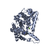
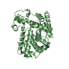
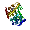
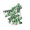
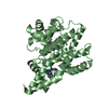
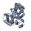
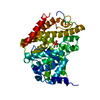
| Title | Crystal structure of human phosphodiesterase 4d with regulatory domain and d155871 | ||||||
 Components Components | cAMP-specific 3',5'-cyclic phosphodiesterase 4D | ||||||
 Keywords Keywords | HYDROLASE / phosphodiesterase / PDE4D / UCR2 / Alternative splicing / cAMP / Cytoplasm / Cytoskeleton / Membrane / Metal-binding / Phosphoprotein | ||||||
| Function / homology |  Function and homology information Function and homology informationsignaling receptor regulator activity / negative regulation of relaxation of cardiac muscle / negative regulation of heart contraction / 3',5'-cyclic-AMP phosphodiesterase / positive regulation of interleukin-5 production / negative regulation of adenylate cyclase-activating G protein-coupled receptor signaling pathway / regulation of cardiac muscle cell contraction / establishment of endothelial barrier / regulation of calcium ion transmembrane transport via high voltage-gated calcium channel / heterocyclic compound binding ...signaling receptor regulator activity / negative regulation of relaxation of cardiac muscle / negative regulation of heart contraction / 3',5'-cyclic-AMP phosphodiesterase / positive regulation of interleukin-5 production / negative regulation of adenylate cyclase-activating G protein-coupled receptor signaling pathway / regulation of cardiac muscle cell contraction / establishment of endothelial barrier / regulation of calcium ion transmembrane transport via high voltage-gated calcium channel / heterocyclic compound binding / beta-2 adrenergic receptor binding / voltage-gated calcium channel complex / adrenergic receptor signaling pathway / cAMP catabolic process / 3',5'-cyclic-nucleotide phosphodiesterase activity / 3',5'-cyclic-GMP phosphodiesterase activity / regulation of cell communication by electrical coupling involved in cardiac conduction / 3',5'-cyclic-AMP phosphodiesterase activity / DARPP-32 events / cAMP binding / positive regulation of heart rate / negative regulation of cAMP/PKA signal transduction / regulation of release of sequestered calcium ion into cytosol by sarcoplasmic reticulum / positive regulation of interleukin-2 production / cellular response to epinephrine stimulus / calcium channel complex / regulation of heart rate / Turbulent (oscillatory, disturbed) flow shear stress activates signaling by PIEZO1 and integrins in endothelial cells / cellular response to cAMP / calcium channel regulator activity / positive regulation of type II interferon production / T cell receptor signaling pathway / ATPase binding / nuclear membrane / scaffold protein binding / G alpha (s) signalling events / transmembrane transporter binding / cilium / apical plasma membrane / centrosome / enzyme binding / nucleoplasm / membrane / metal ion binding / plasma membrane / cytosol Similarity search - Function | ||||||
| Biological species |  Homo sapiens (human) Homo sapiens (human) | ||||||
| Method |  X-RAY DIFFRACTION / SYNCHROTRON / MOLECULAR REPLACEMENT / Resolution: 2.3 Å X-RAY DIFFRACTION / SYNCHROTRON / MOLECULAR REPLACEMENT / Resolution: 2.3 Å | ||||||
 Authors Authors | Staker, B.L. | ||||||
 Citation Citation | Journal: Nat.Biotechnol. / Year: 2010 Title: Design of phosphodiesterase 4D (PDE4D) allosteric modulators for enhancing cognition with improved safety. Authors: Burgin, A.B. / Magnusson, O.T. / Singh, J. / Witte, P. / Staker, B.L. / Bjornsson, J.M. / Thorsteinsdottir, M. / Hrafnsdottir, S. / Hagen, T. / Kiselyov, A.S. / Stewart, L.J. / Gurney, M.E. | ||||||
| History |
|
- Structure visualization
| Structure viewer | Molecule: MolmilJmol/JSmol |
|---|
- Downloads & links
-Download
| PDBx/mmCIF format | 3g4g.cif.gz | 285 KB | Display | PDBx/mmCIF format |
|---|---|---|---|---|
| PDB format | pdb3g4g.ent.gz | 228.5 KB | Display | PDB format |
| PDBx/mmJSON format | 3g4g.json.gz | Tree view | PDBx/mmJSON format | |
| Others |  Other downloads Other downloads |
-Validation report
| Arichive directory | https://data.pdbj.org/pub/pdb/validation_reports/g4/3g4gftp://data.pdbj.org/pub/pdb/validation_reports/g4/3g4g | HTTPS FTP |
|---|
-Related structure data
| Related structure data | 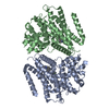 3g45C  3g4iC  3g4kC  3g4lC  3g58C 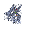 3iadC  3gpz C: citing same article ( |
|---|---|
| Similar structure data |
-Links
 PDBj
PDBj


- Assembly
| Deposited unit | 
| ||||||||
|---|---|---|---|---|---|---|---|---|---|
| 1 | 
| ||||||||
| 2 | 
| ||||||||
| 3 | 
| ||||||||
| 4 | 
| ||||||||
| Unit cell |
| ||||||||
| Details | Authors state that the BIOLOGICAL UNIT IS UNKNOWN. |
-Components
-Protein , 1 types, 4 molecules ABCD
| #1: Protein | Mass: 48370.801 Da / Num. of mol.: 4 / Fragment: residues 299-347 and 360-714 Source method: isolated from a genetically manipulated source Source: (gene. exp.) Homo sapiens (human) / Gene: PDE4D, DPDE3 / Production host:   Spodoptera frugiperda (fall armyworm) Spodoptera frugiperda (fall armyworm)References: UniProt: Q08499, 3',5'-cyclic-nucleotide phosphodiesterase |
|---|
-Non-polymers , 5 types, 183 molecules 


| #2: Chemical | ChemComp-ZN /  Mass: 65.409 Da / Num. of mol.: 4 / Source method: obtained synthetically / Formula: Zn Mass: 65.409 Da / Num. of mol.: 4 / Source method: obtained synthetically / Formula: Zn#3: Chemical | ChemComp-MG /  Mass: 24.305 Da / Num. of mol.: 4 / Source method: obtained synthetically / Formula: Mg Mass: 24.305 Da / Num. of mol.: 4 / Source method: obtained synthetically / Formula: Mg#4: Chemical | ChemComp-CA /  Mass: 40.078 Da / Num. of mol.: 4 / Source method: obtained synthetically / Formula: Ca Mass: 40.078 Da / Num. of mol.: 4 / Source method: obtained synthetically / Formula: Ca#5: Chemical | ChemComp-D71 /  Mass: 375.338 Da / Num. of mol.: 4 / Source method: obtained synthetically / Formula: C19H13N5O4 Mass: 375.338 Da / Num. of mol.: 4 / Source method: obtained synthetically / Formula: C19H13N5O4#6: Water | ChemComp-HOH / | Mass: 18.015 Da / Num. of mol.: 167 / Source method: isolated from a natural source / Formula: H2O |
|---|
-Details
| Sequence details | AUTHORS STATE THAT THE LINKER REGION IS FROM PDE4C THAT WAS SWAPPED TO REPLACE THE NATURAL LINKER, ...AUTHORS STATE THAT THE LINKER REGION IS FROM PDE4C THAT WAS SWAPPED TO REPLACE THE NATURAL LINKER, AS THE NATURAL LINKER WAS HIGHLY CHARGED. |
|---|
-Experimental details
-Experiment
| Experiment | Method: X-RAY DIFFRACTION / Number of used crystals: 1 |
|---|
- Sample preparation
| Crystal | Density Matthews: 2 Å3/Da / Density % sol: 38.36 % |
|---|---|
| Crystal grow | Temperature: 293 K / Method: vapor diffusion, sitting drop / pH: 4.6 Details: 100MM HEPES PH 7.5, 100 MM NACL, 18% PEG 8000, VAPOR DIFFUSION, SITTING DROP, temperature 293K |
-Data collection
| Diffraction | Mean temperature: 100 K |
|---|---|
| Diffraction source | Source: SYNCHROTRON / Site: ALS  / Beamline: 5.0.1 / Wavelength: 0.97 / Beamline: 5.0.1 / Wavelength: 0.97 |
| Radiation | Protocol: SINGLE WAVELENGTH / Monochromatic (M) / Laue (L): M / Scattering type: x-ray |
| Radiation wavelength | Wavelength: 0.97 Å / Relative weight: 1 |
| Reflection | Resolution: 2.3→50 Å / Num. obs: 64589 / % possible obs: 95 % / Redundancy: 2.9 % / Rmerge(I) obs: 0.096 / Net I/σ(I): 6.3 |
| Reflection shell | Resolution: 2.3→2.38 Å / Redundancy: 1.7 % / Rmerge(I) obs: 0.391 / % possible all: 74.3 |
- Processing
| Software |
| ||||||||||||||||||||||||||||||||||||||||||||||||||||||||||||||||||||||||||||||||||||||||||||||||||||||||||||||||||||||||||||||||||||||||||||||||||||||||||||||||||||||||||
|---|---|---|---|---|---|---|---|---|---|---|---|---|---|---|---|---|---|---|---|---|---|---|---|---|---|---|---|---|---|---|---|---|---|---|---|---|---|---|---|---|---|---|---|---|---|---|---|---|---|---|---|---|---|---|---|---|---|---|---|---|---|---|---|---|---|---|---|---|---|---|---|---|---|---|---|---|---|---|---|---|---|---|---|---|---|---|---|---|---|---|---|---|---|---|---|---|---|---|---|---|---|---|---|---|---|---|---|---|---|---|---|---|---|---|---|---|---|---|---|---|---|---|---|---|---|---|---|---|---|---|---|---|---|---|---|---|---|---|---|---|---|---|---|---|---|---|---|---|---|---|---|---|---|---|---|---|---|---|---|---|---|---|---|---|---|---|---|---|---|---|---|
| Refinement | Method to determine structure: MOLECULAR REPLACEMENT / Resolution: 2.3→48.8 Å / Cor.coef. Fo:Fc: 0.942 / Cor.coef. Fo:Fc free: 0.892 / SU B: 7.299 / SU ML: 0.179 / Cross valid method: THROUGHOUT / σ(F): 0 / ESU R: 0.368 / ESU R Free: 0.261 / Stereochemistry target values: MAXIMUM LIKELIHOOD / Details: HYDROGENS HAVE BEEN ADDED IN THE RIDING POSITIONS
| ||||||||||||||||||||||||||||||||||||||||||||||||||||||||||||||||||||||||||||||||||||||||||||||||||||||||||||||||||||||||||||||||||||||||||||||||||||||||||||||||||||||||||
| Solvent computation | Ion probe radii: 0.8 Å / Shrinkage radii: 0.8 Å / VDW probe radii: 1.2 Å / Solvent model: MASK | ||||||||||||||||||||||||||||||||||||||||||||||||||||||||||||||||||||||||||||||||||||||||||||||||||||||||||||||||||||||||||||||||||||||||||||||||||||||||||||||||||||||||||
| Displacement parameters | Biso mean: 22.14 Å2
| ||||||||||||||||||||||||||||||||||||||||||||||||||||||||||||||||||||||||||||||||||||||||||||||||||||||||||||||||||||||||||||||||||||||||||||||||||||||||||||||||||||||||||
| Refinement step | Cycle: LAST / Resolution: 2.3→48.8 Å
| ||||||||||||||||||||||||||||||||||||||||||||||||||||||||||||||||||||||||||||||||||||||||||||||||||||||||||||||||||||||||||||||||||||||||||||||||||||||||||||||||||||||||||
| Refine LS restraints |
| ||||||||||||||||||||||||||||||||||||||||||||||||||||||||||||||||||||||||||||||||||||||||||||||||||||||||||||||||||||||||||||||||||||||||||||||||||||||||||||||||||||||||||
| LS refinement shell | Resolution: 2.3→2.36 Å / Total num. of bins used: 20
|
