 Movie
Movie Controller


+ Open data

- Basic information
| Entry | Database: PDB / ID: 2a72 | ||||||
|---|---|---|---|---|---|---|---|
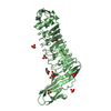
| Title | Structure of the regulator of G-protein signaling domain of RGS7 | ||||||
 Components Components | Regulator of G-protein signalling 7 | ||||||
 Keywords Keywords | SIGNALING PROTEIN / Human RGS7 / regulator of G-protein signaling 7 / GTPase-activating proteins (GAP) / Structural Genomics / Structural Genomics Consortium / SGC | ||||||
| Function / homology |  Function and homology information Function and homology informationdendrite terminus / cell tip / negative regulation of G protein-coupled receptor signaling pathway / regulation of postsynaptic membrane potential / positive regulation of GTPase activity / G-protein alpha-subunit binding / GTPase activator activity / Cooperation of PDCL (PhLP1) and TRiC/CCT in G-protein beta folding / G-protein beta-subunit binding / presynaptic membrane ...dendrite terminus / cell tip / negative regulation of G protein-coupled receptor signaling pathway / regulation of postsynaptic membrane potential / positive regulation of GTPase activity / G-protein alpha-subunit binding / GTPase activator activity / Cooperation of PDCL (PhLP1) and TRiC/CCT in G-protein beta folding / G-protein beta-subunit binding / presynaptic membrane / G alpha (i) signalling events / postsynaptic membrane / neuron projection / intracellular signal transduction / G protein-coupled receptor signaling pathway / GTPase activity / glutamatergic synapse / nucleus / plasma membrane / cytoplasm / cytosol Similarity search - Function | ||||||
| Biological species |  Homo sapiens (human) Homo sapiens (human) | ||||||
| Method |  X-RAY DIFFRACTION / MOLECULAR REPLACEMENT / Resolution: 2 Å X-RAY DIFFRACTION / MOLECULAR REPLACEMENT / Resolution: 2 Å | ||||||
 Authors Authors | Schoch, G.A. / Johansson, C. / Phillips, C. / Debreczeni, J. / Smee, C. / Elkins, J.M. / Sundstrom, M. / Edwards, A. / Arrowsmith, C. / von Delft, F. ...Schoch, G.A. / Johansson, C. / Phillips, C. / Debreczeni, J. / Smee, C. / Elkins, J.M. / Sundstrom, M. / Edwards, A. / Arrowsmith, C. / von Delft, F. / Gileadi, O. / Doyle, D.A. / Structural Genomics Consortium (SGC) | ||||||
 Citation Citation | Journal: Proc.Natl.Acad.Sci.Usa / Year: 2008 Title: Structural diversity in the RGS domain and its interaction with heterotrimeric G protein alpha-subunits. Authors: Soundararajan, M. / Willard, F.S. / Kimple, A.J. / Turnbull, A.P. / Ball, L.J. / Schoch, G.A. / Gileadi, C. / Fedorov, O.Y. / Dowler, E.F. / Higman, V.A. / Hutsell, S.Q. / Sundstrom, M. / ...Authors: Soundararajan, M. / Willard, F.S. / Kimple, A.J. / Turnbull, A.P. / Ball, L.J. / Schoch, G.A. / Gileadi, C. / Fedorov, O.Y. / Dowler, E.F. / Higman, V.A. / Hutsell, S.Q. / Sundstrom, M. / Doyle, D.A. / Siderovski, D.P. | ||||||
| History |
| ||||||
| Remark 300 | BIOMOLECULE: Two biological units are present in the asymmetric unit that are formed from the ...BIOMOLECULE: Two biological units are present in the asymmetric unit that are formed from the combinations of residues 320 to 414 of chain A with 415 to 450 of chain B and residues 320 to 414 of chain B with 415 to 450 of chain A. |
- Structure visualization
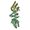
| Structure viewer | Molecule: MolmilJmol/JSmol |
|---|
- Downloads & links
-Download
| PDBx/mmCIF format | 2a72.cif.gz | 72.2 KB | Display | PDBx/mmCIF format |
|---|---|---|---|---|
| PDB format | pdb2a72.ent.gz | 54.1 KB | Display | PDB format |
| PDBx/mmJSON format | 2a72.json.gz | Tree view | PDBx/mmJSON format | |
| Others |  Other downloads Other downloads |
-Validation report
| Arichive directory | https://data.pdbj.org/pub/pdb/validation_reports/a7/2a72ftp://data.pdbj.org/pub/pdb/validation_reports/a7/2a72 | HTTPS FTP |
|---|
-Related structure data
| Related structure data | 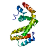 1zv4C  2af0C 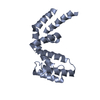 2bt2C 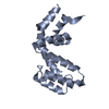 2bv1C 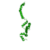 2es0C 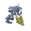 2gtpC 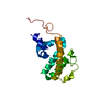 2i59C 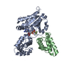 2ihbC 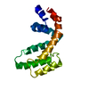 2ihdC 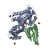 2ik8C 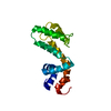 2jm5C 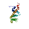 2jnuC 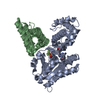 2odeC 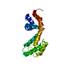 2owiC C: citing same article ( |
|---|---|
| Similar structure data |


-Links
 PDBj
PDBj


- Assembly
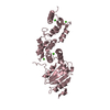
| Deposited unit | 
| ||||||||||||||||||
|---|---|---|---|---|---|---|---|---|---|---|---|---|---|---|---|---|---|---|---|
| 1 |
| ||||||||||||||||||
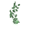
| Unit cell |
| ||||||||||||||||||
| Noncrystallographic symmetry (NCS) | NCS domain:
NCS domain segments: Component-ID: 1 / Ens-ID: 1 / Beg auth comp-ID: ARG / Beg label comp-ID: ARG / End auth comp-ID: ALA / End label comp-ID: ALA / Refine code: 5 / Auth seq-ID: 326 - 443 / Label seq-ID: 9 - 126
|
-Components
| #1: Protein | Mass: 17138.375 Da / Num. of mol.: 2 / Fragment: RESIDUES 320-463 Source method: isolated from a genetically manipulated source Source: (gene. exp.) Homo sapiens (human) / Plasmid: pLIC-SGC / Production host:  #2: Chemical | ChemComp-CL / |   Mass: 35.453 Da / Num. of mol.: 1 / Source method: obtained synthetically / Formula: Cl Mass: 35.453 Da / Num. of mol.: 1 / Source method: obtained synthetically / Formula: Cl#3: Water | ChemComp-HOH / |  Mass: 18.015 Da / Num. of mol.: 231 / Source method: isolated from a natural source / Formula: H2O Mass: 18.015 Da / Num. of mol.: 231 / Source method: isolated from a natural source / Formula: H2O |
|---|
-Experimental details
-Experiment
| Experiment | Method: X-RAY DIFFRACTION / Number of used crystals: 1 |
|---|
- Sample preparation
| Crystal | Density Matthews: 2.1 Å3/Da / Density % sol: 41.1 % |
|---|---|
| Crystal grow | Temperature: 293 K / Method: vapor diffusion, sitting drop / pH: 5.5 Details: PEG 3350, (NH4)2SO4, BIS-TRIS, pH 5.5, VAPOR DIFFUSION, SITTING DROP, temperature 293K |
-Data collection
| Diffraction | Mean temperature: 100 K |
|---|---|
| Diffraction source | Source: ROTATING ANODE / Type: RIGAKU FR-E DW / Wavelength: 1.5418 |
| Detector | Type: RIGAKU RAXIS HTC / Detector: IMAGE PLATE / Date: Jun 13, 2005 |
| Radiation | Protocol: SINGLE WAVELENGTH / Monochromatic (M) / Laue (L): M / Scattering type: x-ray |
| Radiation wavelength | Wavelength: 1.5418 Å / Relative weight: 1 |
| Reflection | Resolution: 2→45.93 Å / Num. all: 19896 / Num. obs: 19896 / % possible obs: 95.9 % / Observed criterion σ(F): 0 / Observed criterion σ(I): 0 / Redundancy: 3.6 % / Biso Wilson estimate: 28.3 Å2 / Rmerge(I) obs: 0.068 / Rsym value: 0.079 / Net I/σ(I): 14.9 |
| Reflection shell | Resolution: 2→2.11 Å / Redundancy: 2.8 % / Rmerge(I) obs: 0.434 / Mean I/σ(I) obs: 2.2 / Num. unique all: 2457 / Rsym value: 0.536 / % possible all: 81.2 |
- Processing
| Software |
| ||||||||||||||||||||||||||||||||||||||||||||||||||||||||||||||||||||||||||||||||||||||||||||||||||||||||||||||||||||||||||||||||||||||||||||||||||||||||||||||||||||||||||
|---|---|---|---|---|---|---|---|---|---|---|---|---|---|---|---|---|---|---|---|---|---|---|---|---|---|---|---|---|---|---|---|---|---|---|---|---|---|---|---|---|---|---|---|---|---|---|---|---|---|---|---|---|---|---|---|---|---|---|---|---|---|---|---|---|---|---|---|---|---|---|---|---|---|---|---|---|---|---|---|---|---|---|---|---|---|---|---|---|---|---|---|---|---|---|---|---|---|---|---|---|---|---|---|---|---|---|---|---|---|---|---|---|---|---|---|---|---|---|---|---|---|---|---|---|---|---|---|---|---|---|---|---|---|---|---|---|---|---|---|---|---|---|---|---|---|---|---|---|---|---|---|---|---|---|---|---|---|---|---|---|---|---|---|---|---|---|---|---|---|---|---|
| Refinement | Method to determine structure: MOLECULAR REPLACEMENT Starting model: 1FQJ_B.pdb Resolution: 2→53 Å / Cor.coef. Fo:Fc: 0.954 / Cor.coef. Fo:Fc free: 0.911 / SU B: 9.269 / SU ML: 0.136 / TLS residual ADP flag: LIKELY RESIDUAL / Cross valid method: THROUGHOUT / σ(F): 0 / σ(I): 0 / ESU R: 0.195 / ESU R Free: 0.184 / Stereochemistry target values: MAXIMUM LIKELIHOOD / Details: HYDROGENS HAVE BEEN ADDED IN THE RIDING POSITIONS
| ||||||||||||||||||||||||||||||||||||||||||||||||||||||||||||||||||||||||||||||||||||||||||||||||||||||||||||||||||||||||||||||||||||||||||||||||||||||||||||||||||||||||||
| Solvent computation | Ion probe radii: 0.8 Å / Shrinkage radii: 0.8 Å / VDW probe radii: 1.2 Å / Solvent model: BABINET MODEL WITH MASK | ||||||||||||||||||||||||||||||||||||||||||||||||||||||||||||||||||||||||||||||||||||||||||||||||||||||||||||||||||||||||||||||||||||||||||||||||||||||||||||||||||||||||||
| Displacement parameters | Biso mean: 28.8 Å2
| ||||||||||||||||||||||||||||||||||||||||||||||||||||||||||||||||||||||||||||||||||||||||||||||||||||||||||||||||||||||||||||||||||||||||||||||||||||||||||||||||||||||||||
| Refinement step | Cycle: LAST / Resolution: 2→53 Å
| ||||||||||||||||||||||||||||||||||||||||||||||||||||||||||||||||||||||||||||||||||||||||||||||||||||||||||||||||||||||||||||||||||||||||||||||||||||||||||||||||||||||||||
| Refine LS restraints |
| ||||||||||||||||||||||||||||||||||||||||||||||||||||||||||||||||||||||||||||||||||||||||||||||||||||||||||||||||||||||||||||||||||||||||||||||||||||||||||||||||||||||||||
| Refine LS restraints NCS | Dom-ID: 1 / Auth asym-ID: A / Ens-ID: 1 / Refine-ID: X-RAY DIFFRACTION
| ||||||||||||||||||||||||||||||||||||||||||||||||||||||||||||||||||||||||||||||||||||||||||||||||||||||||||||||||||||||||||||||||||||||||||||||||||||||||||||||||||||||||||
| LS refinement shell | Resolution: 2→2.052 Å / Total num. of bins used: 20
| ||||||||||||||||||||||||||||||||||||||||||||||||||||||||||||||||||||||||||||||||||||||||||||||||||||||||||||||||||||||||||||||||||||||||||||||||||||||||||||||||||||||||||
| Refinement TLS params. | Method: refined / Refine-ID: X-RAY DIFFRACTION
| ||||||||||||||||||||||||||||||||||||||||||||||||||||||||||||||||||||||||||||||||||||||||||||||||||||||||||||||||||||||||||||||||||||||||||||||||||||||||||||||||||||||||||
| Refinement TLS group |
|
