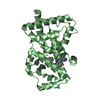
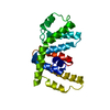
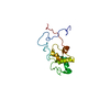
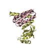
cytosolic small ribosomal subunit / rRNA binding / structural constituent of ribosome / translation Similarity search - Function
Ribosomal protein S4, central domain / RNA-binding S4 domain / SH3 type barrels. - #30 / Structural Genomics Hypothetical 15.5 Kd Protein In mrcA-pckA Intergenic Region; Chain A / Ribosomal protein S4, KOW domain / Ribosomal protein S4e / Ribosomal protein S4e, central region / Ribosomal protein S4e, central domain superfamily / Ribosomal family S4e / SH3 type barrels. ...Ribosomal protein S4, central domain / RNA-binding S4 domain / SH3 type barrels. - #30 / Structural Genomics Hypothetical 15.5 Kd Protein In mrcA-pckA Intergenic Region; Chain A / Ribosomal protein S4, KOW domain / Ribosomal protein S4e / Ribosomal protein S4e, central region / Ribosomal protein S4e, central domain superfamily / Ribosomal family S4e / SH3 type barrels. / OB fold (Dihydrolipoamide Acetyltransferase, E2P) / S4 RNA-binding domain profile. / S4 RNA-binding domain / S4 domain / RNA-binding S4 domain / RNA-binding S4 domain superfamily / Roll / Ribosomal protein L2, domain 2 / Roll / Beta Barrel / Mainly Beta / Alpha Beta Similarity search - Domain/homology
Resolution: 1.75→1.78 Å / Redundancy: 11.2 % / Rmerge(I) obs: 0.433 / Mean I/σ(I) obs: 9.3 / Num. unique all: 2035 / % possible all: 100
-
Processing
Software
Name
Version
Classification
HKL-2000
datacollection
SnB
phasing
RESOLVE
modelbuilding
PHENIX
(phenix.refine: 1.5_2)
refinement
HKL-2000
datareduction
HKL-2000
datascaling
RESOLVE
phasing
Refinement
Method to determine structure: SAD / Resolution: 1.75→28.921 Å / SU ML: 0.2 / Cross valid method: THROUGHOUT / σ(F): 1.04 / Stereochemistry target values: ML
Rfactor
Num. reflection
% reflection
Selection details
Rfree
0.2172
2080
5.15 %
RANDOM
Rwork
0.1844
-
-
-
obs
0.1861
40427
99.73 %
-
Solvent computation
Shrinkage radii: 0.9 Å / VDW probe radii: 1.11 Å / Solvent model: FLAT BULK SOLVENT MODEL / Bsol: 48.632 Å2 / ksol: 0.351 e/Å3
Refinement step
Cycle: LAST / Resolution: 1.75→28.921 Å
Protein
Nucleic acid
Ligand
Solvent
Total
Num. atoms
1473
0
0
193
1666
Refine LS restraints
Refine-ID
Type
Dev ideal
Number
X-RAY DIFFRACTION
f_bond_d
0.016
1493
X-RAY DIFFRACTION
f_angle_d
1.532
2007
X-RAY DIFFRACTION
f_dihedral_angle_d
14.392
558
X-RAY DIFFRACTION
f_chiral_restr
0.11
238
X-RAY DIFFRACTION
f_plane_restr
0.007
253
LS refinement shell
Resolution (Å)
Rfactor Rfree
Num. reflection Rfree
Rfactor Rwork
Num. reflection Rwork
Refine-ID
% reflection obs (%)
1.75-1.7907
0.2399
128
0.207
2565
X-RAY DIFFRACTION
99
1.7907-1.8355
0.2505
136
0.2063
2521
X-RAY DIFFRACTION
100
1.8355-1.8851
0.2428
133
0.2091
2573
X-RAY DIFFRACTION
100
1.8851-1.9406
0.2501
126
0.1951
2573
X-RAY DIFFRACTION
100
1.9406-2.0032
0.2353
168
0.1875
2553
X-RAY DIFFRACTION
100
2.0032-2.0748
0.2083
136
0.1772
2521
X-RAY DIFFRACTION
100
2.0748-2.1578
0.2565
124
0.1828
2582
X-RAY DIFFRACTION
100
2.1578-2.256
0.1652
125
0.1755
2581
X-RAY DIFFRACTION
100
2.256-2.3749
0.2226
151
0.1685
2530
X-RAY DIFFRACTION
100
2.3749-2.5236
0.188
137
0.1834
2584
X-RAY DIFFRACTION
100
2.5236-2.7183
0.2025
157
0.1829
2532
X-RAY DIFFRACTION
100
2.7183-2.9916
0.1935
169
0.1957
2565
X-RAY DIFFRACTION
100
2.9916-3.4239
0.2818
117
0.1863
2565
X-RAY DIFFRACTION
100
3.4239-4.3115
0.2163
158
0.165
2553
X-RAY DIFFRACTION
100
4.3115-28.9249
0.1863
115
0.1796
2549
X-RAY DIFFRACTION
99
Refinement TLS params.
Method: refined / Origin x: 24.7777 Å / Origin y: 44.7101 Å / Origin z: 13.0267 Å
11
12
13
21
22
23
31
32
33
T
0.1423 Å2
-0.0093 Å2
-0.0136 Å2
-
0.0394 Å2
0.0172 Å2
-
-
0.0826 Å2
L
0.5288 °2
0.0674 °2
0.0759 °2
-
0.3133 °2
0.0969 °2
-
-
0.484 °2
S
-0.0051 Å °
-0.018 Å °
-0.0912 Å °
0.1561 Å °
0.0248 Å °
-0.0062 Å °
-0.0397 Å °
-0.0121 Å °
-0.0139 Å °
Refinement TLS group
Selection details: chain A
+
About Yorodumi
-
News
-
Feb 9, 2022. New format data for meta-information of EMDB entries
New format data for meta-information of EMDB entries
Version 3 of the EMDB header file is now the official format.
The previous official version 1.9 will be removed from the archive.
In the structure databanks used in Yorodumi, some data are registered as the other names, "COVID-19 virus" and "2019-nCoV". Here are the details of the virus and the list of structure data.
Jan 31, 2019. EMDB accession codes are about to change! (news from PDBe EMDB page)
EMDB accession codes are about to change! (news from PDBe EMDB page)
The allocation of 4 digits for EMDB accession codes will soon come to an end. Whilst these codes will remain in use, new EMDB accession codes will include an additional digit and will expand incrementally as the available range of codes is exhausted. The current 4-digit format prefixed with “EMD-” (i.e. EMD-XXXX) will advance to a 5-digit format (i.e. EMD-XXXXX), and so on. It is currently estimated that the 4-digit codes will be depleted around Spring 2019, at which point the 5-digit format will come into force.
The EM Navigator/Yorodumi systems omit the EMD- prefix.
Related info.:Q: What is EMD? / ID/Accession-code notation in Yorodumi/EM Navigator
Yorodumi is a browser for structure data from EMDB, PDB, SASBDB, etc.
This page is also the successor to EM Navigator detail page, and also detail information page/front-end page for Omokage search.
The word "yorodu" (or yorozu) is an old Japanese word meaning "ten thousand". "mi" (miru) is to see.
Related info.:EMDB / PDB / SASBDB / Comparison of 3 databanks / Yorodumi Search / Aug 31, 2016. New EM Navigator & Yorodumi / Yorodumi Papers / Jmol/JSmol / Function and homology information / Changes in new EM Navigator and Yorodumi
 Movie
Movie Controller

 Yorodumi
 Open data
 Basic information
 Components
 Keywords
 Function and homology information
 Thermoplasma acidophilum (acidophilic)
 X-RAY DIFFRACTION /
  Authors
 Citation
 Structure visualization
 Downloads & links
 Other downloads


 PDBj
PDBj
 Assembly


 Mass: 18.015 Da / Num. of mol.: 193 / Source method: isolated from a natural source / Formula: H2O
 Sample preparation
 / Beamline: X4A / Wavelength: 0.97912 Å
 Processing